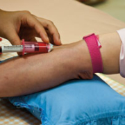
Advances in prenatal testing based on cell free fetal DNA in maternal plasma
Non-invasive prenatal testing (NIPT) based on cell free fetal DNA (cffDNA) circulating in maternal blood is moving rapidly forward. With promises of improved safety, earlier detection and easier access to tests, NIPT has the potential to bring many positive benefits to prenatal care. Here we discuss the recent developments in this area.
by Dr Melissa Hill, Dr Angela Barrett, Dr Helen White and Professor Lyn Chitty
Non-invasive testing using cell free fetal DNA
Prenatal diagnosis of genetic conditions or aneuploidy has traditionally required invasive diagnostic tests [chorionic villus sampling (CVS) and amniocentesis] which carry a small but significant risk of miscarriage of around 1% and can only be safely conducted after 11 weeks in pregnancy. In 1997 Lo and colleagues identified the presence of cell free fetal DNA (cffDNA) in maternal plasma and in doing so opened the door to a safer approach to prenatal diagnosis whereby non-invasive prenatal testing (NIPT) of fetal genetic material could be performed using a maternal blood test [1].
The cffDNA is an attractive target for prenatal testing. In addition to avoiding the risk of miscarriage it is anticipated that NIPT will be available early in pregnancy as cffDNA can be detected from 4 to 5 weeks with sufficient levels for analysis by 7 to 9 weeks. The cffDNA emanates from trophoblast cells in the placenta and is pregnancy specific as it is cleared from the circulation within 30 minutes of delivery. It is now also evident that the whole fetal genome is represented in the maternal plasma, suggesting that tests for many genetic conditions will be possible [2].
The major barrier to developing specific prenatal tests based on cffDNA has been the relative concentration of the fetal material. The cffDNA represents only a small proportion (around 10%) of the cell free DNA that is present in the maternal circulation, as the vast majority is maternal in origin. As a result, it is difficult to determine what genetic information is specific to the fetus against the large background of maternal cell free DNA.
For this reason NIPT was initially limited to the identification of alleles present in the fetus but not in the mother because they were inherited from the father or because they arose de novo. These tests include fetal sex determination, which uses targets on the Y chromosome, fetal rhesus genotyping in Rhesus D (RhD) negative mothers, and paternally inherited or de novo autosomal dominant single gene disorders. All of which can be conducted using relatively straightforward molecular techniques. More recently, new technologies such as digital PCR and massively parallel sequencing (MPS) have allowed researchers to develop NIPT for single gene disorders where parents have the same mutations and for aneuploidies, both of which need to take into account the presence of the mother’s allele or chromosomes.
Early clinical successes with non-invasive testing
The two early success stories for NIPT have been fetal rhesus genotyping and fetal sex determination, which are performed using real-time quantitative PCR. Analysis of cffDNA in the plasma of RhD-negative pregnant women who have a past history of haemolytic disease of the newborn or have elevated levels of Anti-D antibodies has been used clinically to determine the fetal RHD status for almost a decade. Large scale validation studies demonstrate high specificity and sensitivity and fetal RHD typing of all RhD-negative pregnant women has the potential to become routine clinical practice in the next few years.
Similarly, NIPT for fetal sex determination is increasingly offered as standard genetic care for women with pregnancies at risk of genetic conditions that primarily affect a particular sex. Fetal sex determination informs the need for genetic diagnosis, allowing up to 50% of carriers to avoid unnecessary invasive testing, and is important for guiding pregnancy management in some conditions. The test has been shown to be reliable when performed after 7 weeks gestation [3], and clinical utility was demonstrated in a recent UK audit as only 32.9% of women subsequently underwent invasive testing [4]. Importantly for implementation, NIPT is viewed positively by women who have had the test and has been shown to be cost neutral compared to invasive testing, which means women can have the clinical benefits of the test at no extra cost to health services [4].
NIPT for single gene disorders
The first use of cffDNA for the diagnosis of a single gene disorder was for the autosomal dominant condition myotonic dystrophy [5]. NIPT has also been possible for other paternally inherited autosomal dominant disorders such as early onset primary dystonia and Huntington’s disease. NIPT for the autosomal dominant condition achondroplasia, which commonly occurs as a de novo mutation, has also been successful. NIPT for autosomal recessive or maternally transmitted autosomal dominant disorders is more difficult due to the need to distinguish between the maternal and fetal free DNA. Exclusion of the paternal mutation is possible in autosomal recessive conditions where the parents carry different mutations. If the paternal allele is detected, there is a 50% risk that the fetus will inherit the disorder, and invasive testing would be recommended. If the paternal allele is not detected invasive testing is not required.
It is now clear that NIPT can be successfully applied to recessive conditions where parents carry the same mutation using digital PCR, which allows high copy number counting and quantification of alleles. Digital PCR requires the dilution of template DNA to an average concentration of less than one molecule per well, and hundreds to thousands of replicates of a PCR reaction are analysed. The mutation status of the fetus is predicted with an approach known as relative mutation dosage (RMD), which is based on the premise that if both the woman and the fetus are heterozygous there will be an allelic balance between the wild type and normal alleles; if the fetus is homozygous for either the mutant or the wild type allele there will be an over-representation of one or the other [6]. Another approach to identify single gene disorders non-invasively that we may see more of in the future is MPS, which has been used to determine the inheritance of two different β-thalassemia alleles [2].
NIPT for aneuploidies
Prenatal screening and diagnosis for fetal aneuploidy is offered routinely to all pregnant women in many countries to detect trisomy 21 (Down’s syndrome), trisomy 18 (Edwards syndrome) and trisomy 13 (Patau syndrome). The extensive scale of current prenatal screening programmes for these conditions means that success in developing accurate NIPT for aneuploidies will transform antenatal care. Several approaches have been explored including fetal specific epigenetic markers and SNP based methods. The most promising approach to date utilises MPS, which allows large scale single molecule counting to detect the increase in the number of sequences that result from the trisomic chromosome.
The first successful proof-of-principle studies utilising MPS to detect fetal aneuploidy from maternal plasma were published in 2008 [7,8]. Using this methodology, millions of short DNA sequences are generated from genomic locations. The sequences are then compared with the known human genome sequence, to establish how many sequences have been derived from each chromosome. For example, by comparing the total number of uniquely mapped chromosome 21 sequences obtained from a cfDNA sample with the number obtained from a normal genomic DNA sample, very small increases in the amount of chromosome 21 can be detected in the cfDNA sample if the fetus carries an additional chromosome 21.
Several large scale validation studies have subsequently demonstrated high levels of sensitivity (100%) and specificity (98–99%) using MPS. Efforts to decrease costs and increase throughput have seen many groups use multiplexing of patient samples into samples libraries that are run on one lane of the sequencing platform (2–12 patients per lane). Another strategy to decrease costs has been the use of targeted or ‘chromosome-selective’ MPS approaches where the sequencing assay is targeted to non-polymorphic loci on specific chromosomes such as 18 and 21 [10]. Studies with targeted MPS also show the potential for greater accuracy with the use of a novel bioinformatic algorithim (FORTE) that considers the proportion of specific cffDNA in the samples and accounts for the prior risk of trisomy (taken from published data on maternal and fetal gestational age related risks) to predict the likelihood of fetal trisomy for each patient [9].
Following the success of the validation studies, NIPT for aneuploidy is being offered through commercial providers in some countries (Sequenom, BGI, Berry Genomics, Aria, LifeCodexx). It is not yet clear, however, how these new tests will be introduced more widely into antenatal care. At present the small false positive rate means that the test is considered an “advanced screening test” that should be confirmed by invasive testing. Other considerations for implementation include the cost of the technology, the gestational limits of the test and the structure of existing screening programmes. All of these factors will impact on whether NIPT is introduced as a replacement for invasive testing or as an adjunct to current screening tests offered to all women.
Conclusions
NIPT is rapidly bringing about dramatic changes to antenatal care. Fetal sex determination and RhD genotyping are now available as clinical services in a number of countries. Testing is already possible for some single gene disorders, and new technologies such as digital PCR and MPS are allowing the challenges of testing for recessive conditions and aneuploidies to be met. Successful implementation, however, will require more than the development of laboratory tests and we must consider ethical issues, research stakeholder views and assess implementation strategies to ensure NIPT is offered in a way that best meets women’s needs. For this reason studies such as the RAPID programme in the UK (www.rapid.nhs.uk) that look at all aspects of test development and implementation are important.
Notification
This article summarises a recent review published in Best Practice in Clinical and Obstetric Gynaecology: Hill M, Barrett AN, White H, Chitty LS. Uses of cell free fetal DNA in maternal circulation. Best Pract Res Clin Obstet Gynaecol. 2012 Apr 27 [Epub ahead of print].
References
1. Lo YM, Corbetta N, Chamberlain PF, et al. Presence of fetal DNA in maternal plasma and serum. Lancet. 1997; 350: 485–487.
2. Lo Y, Chan K, Sun H, et al. Maternal plasma DNA sequencing reveals the genome-wide genetic and mutational profile of the fetus. Sci Transl Med. 2010; 2: 61ra91.
3. Devaney SA, Palomaki GE, Scott JA, et al. Noninvasive fetal sex determination using cell-free fetal DNA: a systematic review and meta-analysis. JAMA. 2011; 306: 627–636.
4. Hill M, Lewis C, Jenkins L, Allen S, Elles R, Chitty LS. Implementing non-invasive prenatal fetal sex determination using cell free fetal DNA in the United Kingdom. Expet Opin Biol Ther. 2012; Suppl 1: S119–126.
5. Amicucci P, Gennarelli M, Novelli G, et al. Prenatal diagnosis of myotonic dystrophy using fetal DNA obtained from maternal plasma. Clin Chem. 2000; 46: 301–302.
6. Lun FM, Tsui NB, Chan KC, et al. Noninvasive prenatal diagnosis of monogenic diseases by digital size selection and relative mutation dosage on DNA in maternal plasma. Proc Natl Acad Sci U S A. 2008; 105: 19920–19925.
7. Fan HC, Blumenfeld YJ, Chitkara U, et al. Noninvasive diagnosis of fetal aneuploidy by shotgun sequencing DNA from maternal blood. Proc Natl Acad Sci U S A. 2008; 105: 16266–16271.
8. Chiu RW, Chan KC, Gao Y, et al. Noninvasive prenatal diagnosis of fetal chromosomal aneuploidy by massively parallel genomic sequencing of DNA in maternal plasma. Proc Natl Acad Sci U S A. 2008; 105: 20458–20463.
9. Sparks AB, Struble CA, Wang ET, et al. Optimized Non-invasive evaluation of fetal aneuploidy risk using cell-free DNA from maternal blood. Am J Obstet Gynecol. 2012; 206: 319.
The authors
Melissa Hill PhD1, Angela Barrett PhD1,
Lyn Chitty MRCOG, PhD1* and
Helen White PhD2
1 Clinical and Molecular Genetics Unit, UCL Institute of Child Health and Great Ormond Street Hospital for Children NHS Foundation Trust, London, UK
2 National Genetics Reference Laboratory (Wessex), Salisbury District Hospital, Salisbury, UK
*Corresponding author
e-mail: l.chitty@ucl.ac.uk


