Bio-Rad - Preparing for a Stress-free QC Audit

Getting the most from every drop – new advances in blood separation

Getting the most from every drop – new advances in blood separation

Alexandra Sommer, Senior Product Manager, Clinical Diagnostics at Tecan

Liquid biopsies are poised to play a central role in the future of diagnostics, especially in oncology.1 Currently, a conventional tissue biopsy is the gold standard in the diagnosis of many malignancies, but this technique comes with many drawbacks, including its invasive nature, the risk to patients, the need to fix the samples, and the difficulty in applying it to all cancer types and patients. Liquid biopsy allows genomic and proteomic assessment of components in the blood, or other sampled fluids, as an alternative, less invasive and more convenient method of monitoring cancer patients during and after treatment. Following sampling, the buffy coat and plasma can be separated from the erythrocytes by centrifugation, and analysed to understand the health status of the patient, and also check sample quality. Individual biomarkers in both the plasma and buffy coat – including circulating tumour cells (CTCs), circulating tumour DNA and circulating tumour ribonucleic acid (ctDNA, ctRNA), and exosomes – can be analysed to provide further insights into a patient’s cancer, offering a comprehensive view of tumour heterogeneity, and giving an overview of tumour characteristics such as progression, staging, heterogeneity, gene mutations, and clonal evolution. This can assist clinicians with devising personalised therapeutic regimens, as well as allowing continuous monitoring – by repeated sampling – to measure treatment efficacy and screen for therapeutic resistance.

CTCs
CTCs are intact tumour cells that are released into the bloodstream from primary or secondary tumour sites, and their migration through the circulatory system results in the development of distant metastases. They can be collected in the buffy coat following blood separation by centrifugation, and analysis of CTCs provides diagnostic and prognostic information in a number of cancer types. Their detection and characterisation is an important tool for monitoring and preventing the development of overt metastatic disease, and CTCs have also shown potential as screening biomarkers for early detection of cancers.2

ctDNA
Cancer cells shed naked DNA molecules, known as ctDNA, into the bloodstream. This genetic material can be isolated from plasma, and has become one of the main markers to investigate the mutational status of cancers through the use of next generation sequencing. Comparison of ctDNA with cfDNA – a normal product of cell turnover that is detectable in healthy individuals – can be used to identify mutations in tumours are not an exact match to an individual’s DNA, providing highly specific markers for various cancers.3 Recent advances in precision medicine have also allowed analysis of ctDNA to guide treatment decisions, predicting response and resistance to targeted therapies and immunotherapies. While most current clinical practice using ctDNA has been aimed at identifying druggable and resistance mutations for directing systemic therapy decisions, newer research is evaluating its potential as a marker of minimal residual disease, and as a useful screening tool to detect cancers in the general population.4

Exosomes
Exosomes are nano-vesicles released by various cell types, and carry a variety of ‘molecular messengers’ – including proteins, RNAs, DNAs and lipids. There is substantial evidence that exosomes are involved in intercellular communication by exchanging these messenger molecules between cells, and that they play important roles in cancer development. They can be isolated from plasma following blood separation, and recent studies have shown that exosomes are superior to CTCs and ctDNA for early diagnosis, disease monitoring, and prognostic prediction. However, they are more challenging to extract and study than other blood constituents, so improved strategies to isolate exosomes from bodily fluids – and to profile exosomal contents in a fast and sensitive way – are required to allow the practical application of exosome-based liquid biopsies for precision cancer medicine.5

Liquid biopsy applications also go beyond cancer. For example, non-invasive prenatal testing (NIPT) is becoming a preferred choice for many mothers undergoing testing for chromosomal abnormalities, as a safer alternative to amniocentesis. This is an increasingly important consideration as the average age of mothers continues to increase,6 prompting significant research interest in this area. Liquid biopsy approaches are also showing promise for monitoring patients following organ transplant. For example, a number of biomarkers – including donor-derived cell-free DNA, microRNAs and exosomes – are showing high potential for determining the long-term prognosis for kidney transplantation patients.7

The challenge of separating blood samples
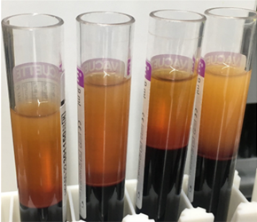
Blood collected for liquid biopsy must be separated into its constituents – plasma, buffy coat and erythrocytes – in order to study the markers described above. Traditional manual separation techniques are slow, and are susceptible to human error, even when performed by an experienced technician. Following centrifugation, the delicate buffy coat forms a thin layer between the plasma and erythrocytes, and manual isolation – usually carried out with a Pasteur pipette – carries the risk of contamination between the phases. Even when automation technology is available, it can be challenging to identify liquid-liquid interfaces, because barcodes and labels may interfere with optical sensors. With the increasing number of applications for liquid biopsies – from cancer monitoring to NIPT – laboratories need ways to streamline the workflow and overcome the bottleneck of blood separation.

A centrifuged blood

 (A) centrifuged blood

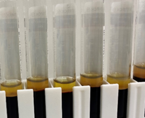
B after plasma separation

(B) after plasma separation

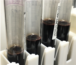
C after buffy separation

(C) after buffy separation

Presenting Phase Separator™

Phase Separator from Tecan is an advancement in liquid-liquid separation technology that offers precision and speed, helping to streamline workflows and improve efficiency in both research and clinical laboratories. It is a unique, pressure-based detection technology for the Air Flexible Channel Arm™ (Air FCA) on Tecan’s flagship liquid handling platform, the Fluent® Automation Workstation. This novel method addresses the critical challenge of detecting liquid-liquid interfaces and effectively separating neighbouring phases, while avoiding the risk of contamination, making it ideal for removing plasma or buffy coat layers from centrifuged blood samples.

Phase Separator can achieve remarkable speeds, working in either tubes or plates, by combining phase detection with the pipetting action. Processing speed is further enhanced when all eight channels on the Air FCA are used in parallel – enabling the aspiration of plasma from 24 tubes of centrifuged blood in under 10 minutes* – and twice as fast again on dual-arm systems. These benefits come with no loss of bench space, and no additional equipment to maintain. Crucially, the technology detects liquid levels with pinpoint precision inside the tube or plate, so there is no interference from barcodes or other labware markings.

Proven against traditional methods

The value of Phase Separator for sample preparation in pre-natal testing has been investigated, in efforts to optimize the quality and quantity of cfDNA. Donated blood from women in different stages of their pregnancy was collected in different tube types – including Cell-Free DNA BCT® (Streck) and PAXgene® Blood ccfDNA Tube (PreAnalytiX®) – and the plasma was separated using either the standard, laboratory-validated method (labelled PM) or the Phase Separator. Between five and seven millilitres of plasma was separated per sample, based on input volume and tube type.

The Phase Separator was also tested with samples that had undergone a second centrifugation step after plasma separation, to determine the impact on cfDNA extraction, presented as percentage of foetal fraction isolated from total cfDNA in a sample (Figure 2). Different separation conditions using the Phase Separator were tested, and compared with an established and validated manual separation and cfDNA extraction protocol:

  • Increased quality: Aspirate and aliquot plasma to destination tube. Upon phase detection, dispense back 150 µl of plasma from disposable tip, then transfer remaining plasma in the disposable tip to a destination tube for extraction (>5 mm remaining volume)
  • Increased quantity: Aspirate and aliquot plasma to destination tube. Upon phase detection, dispense back 150 µl of plasma from disposable tip and transfer remaining plasma in the disposable tip to a destination tube for extraction. Continue plasma separation after phase detection down to <5 mm remaining plasma volume on top of buffy coat layer.

The percentage of foetal cfDNA isolated correlated well with the stage of pregnancy, with % foetal fraction (FF) in the late pregnancy samples (week 38/39) being higher than the percentage identified in earlier pregnancy samples. Importantly, no differences in the overall percentage of foetal cfDNA isolated from the total cfDNA were detected when using with Phase Separator technology compared to the lab validated method, independent of the tube type, secondary centrifugation or separation conditions. This feasibility study provides supporting data on the quantity and quality of the plasma separated using Phase Separator technology, setting the foundation for further validation studies that could aim to  replace labour intensive manual plasma separation.

Figure 2

Figure 2: Percentage of foetal cfDNA collected across five different donors using various sampling conditions.

On the pulse of innovation

Automating phase separation allows blood samples to be processed quickly and accurately, reducing the likelihood of sample loss due to contamination. This will result in productivity gains and cost savings for laboratories. The technology will be especially important in the rapidly evolving field of liquid biopsies, with benefits for disease diagnostics and monitoring across a range of medical fields.

For further information about Tecan’s Phase Separator, please visit www.tecan.com/phase-separator.

*Timing is volume dependent. Time given for separation volumes of 5 ml.

The Fluent Phase Separator is an open liquid handling platform. No performance claims are made by Tecan. Performance validation is required by the customer.

References

  1. Lone SN, Nisar S, Masood T et al. Liquid biopsy: a step closer to transform diagnosis, prognosis and future of cancer treatments. Molecular Cancer (2022). 21:79. https://doi.org/10.1186/s12943-022-01543-7
  2. Hirahata T, ul Quraish R, ul Quarish A et al. Liquid Biopsy: A Distinctive Approach to the Diagnosis and Prognosis of Cancer. Cancer Informatics (2022). 21:1-7. https://doi.org/10.1177/11769351221076062
  3. Cowling T and Loshak H. An Overview of Liquid Biopsy for Screening and Early Detection of Cancer. CADTH (2019). 179. ISSN: 1488-6324
  4. Dang DK and Park BH. Circulating tumor DNA: current challenges for clinical utility. J Clin Invest (2022). 132(12):e154941. https://doi.org/10.1172/JCI154941
  5. Yu D, Li Y, Wang M. et al.Exosomes as a new frontier of cancer liquid biopsy. Mol Cancer (2022). 21:56. https://doi.org/10.1186/s12943-022-01509-9
  6. Bedei I, Wolter A, Weber A et al. Chances and Challenges of New Genetic Screening Technologies (NIPT) in Prenatal Medicine from a Clinical Perspective: A Narrative Review. Genes (2021). 12:501. https://doi.org/10.3390/genes1204050
  7. Chancharoenthana W, Traitanon O, Leelahavanichkul A et al. Molecular immune monitoring in kidney transplant rejection: a state-of-the art review. Immunol (2023). 14:1206929. https://doi.org/10.3389/fimmu.2023.1206929