MALDI-TOF mass spectrometry for rapid identification and subtyping of H. cinaedi strains isolated from humans and animals
Although Helicobacter cinaedi infection is now recognized as an increasingly important emerging disease in humans, it is difficult to identify particular isolates due to their unusual phenotypic profiles and similarity of 16S rRNA sequences among closely related helicobacters. MALDI-TOF MS resolved the present difficulties associated with the identification of H. cinaedi.
by Takako Taniguchi and Prof. Naoaki Misawa
Helicobacter cinaedi infection
Helicobacter cinaedi, was first recognized as a Campylobacter-like organism (CLO), is a Gram-negative, spiral-shaped, motile, microaerobic bacterium, and is now classified into enterohepatic Helicobacter species [1]. This organism was first isolated from homosexual men and was initially recognized as a rectal and intestinal pathogen among members of that population [1]. The first case of H. cinaedi bacteremia in Japan was in an HIV-negative patient, but was receiving immunosuppressive therapy after renal transplantation [2]. Moreover, a few cases of infection with H. cinaedi isolated from feces and blood from an apparently non-immunocompromised child and adult have been reported [3]. Since then, H. cinaedi has become thought of as an opportunistic pathogen that causes bacteremia, cellulitis, septicemia and enteritis in immunocompromised patients [4, 5], immunocompetent patients and even healthy individuals [6]. Kitamura et al. reported an outbreak of nosocomial H. cinaedi infections caused by direct person-to-person transmission [6]. Therefore, healthcare workers need to pay attention to H. cinaedi infection as an increasingly important emerging disease in humans.
Epidemiology
H. cinaedi-like organisms have also been isolated from non-human sources such as dogs, cats, monkeys, hamsters and other rodents [7–10], suggesting that the organism may be widespread in a broad range of animal species. As Gebhart et al. reported that H. cinaedi was found in 75% of the healthy hamsters used in their study [9], it was hypothesized that hamsters might be an important reservoir for human infection [7, 9]. However, no reliable epidemiological evidence of zoonosis has been demonstrated for human cases of H. cinaedi infection [3].
Diagnosis
To isolate H. cinaedi from blood, blood was usually collected in BACTEC culture bottles and incubated in a BACTEC 9050 blood culture system (Becton Dickinson, BD Biosciences) for at least 5 to 7 days. When the incubation time was less than 5 days or other culture systems were used, the organism was not often isolated. Earlier research suggested that certain patients with H. cinaedi infection may remain undiagnosed or incorrectly diagnosed because of difficulties in detecting the bacteria by conventional culture methods [2].
We previously isolated at least six different spiral-shaped organisms including H. cinaedi and H. bilis in a puppy with bloody diarrhoea [11]. These organisms were identified based on their morphology, biochemical traits, whole-cell protein profiles, and the results of molecular analysis of their 16S rDNA sequences. However, the biochemical identification of Helicobacter strains based on a limited number of tests is difficult because helicobacters frequently exhibit unusual phenotypic profiles, even in the same species [10, 12]. Furthermore, H. cinaedi cannot be clearly discriminated from H. bilis on the basis of 16S rRNA sequences because of the high level of sequence similarity (greater than 98%) [12].
Application of MALDI-TOF MS for rapid identification and subtyping of H. cinaedi strains
Recently, matrix-assisted laser desorption/ionization time-of-flight mass spectrometry (MALDI-TOF MS) has made it possible to analyse the protein composition of a bacterial cell based on intact-cell mass spectrometry (ICMS) profiles as a new technique for species identification. The technique is simple, rapid and accurate for identifying microorganisms regardless of their characteristics, such as Gram-negative and Gram-positive bacteria, mycobacteria, anaerobes and yeast species [13, 14]. MALDI-TOF MS has an advantage in that it has a low-cost performance and is independent of the age of the culture, growth conditions or medium selected, making it applicable for routine bacterial identification in clinical laboratories. Although there is a commercially available library that includes more than three thousand kinds of microorganism such as bacteria, yeast and fungus for identification and phylogenic analysis (MALDI Biotyper Reference Library, Bruker Daltonics), we use a library created in-house.
Therefore, we considered that MALDI-TOF MS might resolve the present difficulties with identification of H. cinaedi. Furthermore, we examined whether H. cinaedi strains isolated from different animals could be differentiated or subtyped by their ICMS profiles [15].
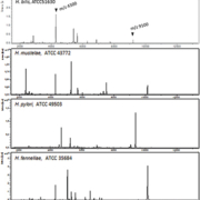
As shown in Figure 1, although common peaks were detected in the H. cinaedi and H. bilis strains examined, the m/z 5200 and 10400 peaks were detected only in strains of H. cinaedi. These peaks showed good reproducibility regardless of the isolate origins, different media and passage numbers on the same medium. Therefore, the ICMS profile of H. cinaedi could be completely differentiated from those of H. bilis. Furthermore, the ICMS profile of H. cinaedi was also distinguishable from those of H. mustelae, H. pylori, H. fennelliae and H. canis, indicating that ICMS profiling using MALDI-TOF MS is applicable for the identification of H. cinaedi.
Cluster analysis of H. cinaedi strains based on the ICMS profiles
Several papers report that direct contact with pets may be a possible route of infection in humans [3–5]; however, details regarding the pathogenesis and epidemiological features, including routes of infection of animal isolates in the context of both humans and animals, are not fully understood. No reliable epidemiological evidence of zoonosis has been demonstrated for human infections caused by H. cinaedi. Therefore, ICMS profiles of H. cinaedi strains isolated from humans and animals were measured, and a phyloproteomic tree was constructed in order to analyse the relationships between the strains. As a result, these H. cinaedi strains were clearly divided into two groups. All of the strains isolated from humans belonged to Cluster 2. All the other animal-derived strains belonged to Cluster 1 (Fig. 2). Interestingly, the ICMS-based phyloproteomic tree agreed with the phylogenetic tree that had been based on the nucleotide sequences of the hsp60 gene. These H. cinaedi strains were also clearly divided into two groups by the hsp60-gene-based phyloproteomic tree. Thus, the data from phyloproteomic and phylogenetic analysis suggest that human strains of H. cinaedi may be distinct from animal strains. Kiehlbauch et al. also reported that there may be subgroups within H. cinaedi isolated from humans, dogs, cats and hamsters that correlate with the host source on the basis of DNA–DNA hybridization and ribotyping analyses [12]. The present study appears to support the hypothesis that H. cinaedi from different host sources may form subgroups, which may prompt a revision of the classification of H. cinaedi.
Conclusion
In conclusion, the construction of ICMS profiles using the MALDI-TOF MS approach may be a useful tool for H. cinaedi identification and subtyping. Further investigations will be required to analyse additional strains from a broader area to confirm whether human strains belong to a distinct subtype of H. cinaedi.
References
1. Quinn TC, Goodell SE, et al. Ann Intern Med. 1984; 101: 187–192.
2. Murakami H, Goto M, et al. J Infect Chemother. 2003; 9: 344–347.
3. Orlicek SL, Welch DF, Kuhls TL. J Clin Microbiol. 1993; 31: 569–571.
4. Kiehlbauch JA, Tauxe RV, et al. Ann Intern Med. 1994; 121: 90–93.
5. Matsumoto T, Goto M, et al. J Clin Microbiol. 2007; 45: 2853–2857.
6. Kitamura T, Kawamura Y, et al. J Clin Microbiol. 2007; 45: 31–38.
7. Comunian LB, Moura SB, et al. Curr Microbiol. 2006; 53: 370–373.
8. Fernandez KR, Hansen LM, et al. J Clin Microbiol. 2002; 40: 1908–1912.
9. Gebhart CJ, Fennell CL, et al. J Clin Microbiol. 1989; 27: 1692–1694.
10. Kiehlbauch JA, Brenner DJ, et al. J Clin Microbiol. 1995; 33: 2940–2947.
11. Misawa N, Kawashima K, et al. Vet Microbiol. 2002; 87: 353–364.
12. Vandamme P, Harrington CS, et al. J Clin Microbiol. 2000; 38:.2261–2266.
13. Saffert RT, Cunningham SA, et al. J Clin Microbiol. 2011; 49: 887–892.
14. Stevenson LG, Drake SK, et al. J Clin Microbiol. 2010; 48: 3482–3486.
15. Taniguchi T, Sekiya A, et al. J Clin Microbiol. 2014; 52: 95–102.
The authors
Takako Taniguchi1 MSc and Naoaki Misawa2* DVM, PhD
1Laboratory of Veterinary Public Health, Department of Veterinary Science, Faculty of Agriculture, University of Miyazaki, Miyazaki, Japan
2Center for Animal Disease Control, University of Miyazaki, Miyazaki, Japan
*Corresponding author
E-mail: a0d901u@cc.miyazaki-u.ac.jp


