MicroRNAs show potential as molecular biomarkers for graft-versus-host disease
Graft-versus-host disease is a serious complication following hematopoietic stem cell transplantation (HSCT), with a high mortality rate. A clearer understanding of the molecular pathogenesis may allow robust biomarker identification and improved therapeutic options. MicroRNAs (miRNAs) are short non-coding regulatory RNAs that are expressed in both tissue and body fluids, and show great potential as clinically translatable biomarkers. Here we discuss the field of miRNA biomarker discovery in the setting of HSCT.
by Dr Rachel E. Crossland and Prof. Anne M. Dickinson
Allogeneic hematopoietic stem cell transplant and graft-versus-host disease
Allogeneic hematopoietic stem cell transplant (allo-HSCT) is a curative treatment for many blood cancers. It is based on the transplant of hematopoietic blood and marrow stem cells from related or unrelated donors, and over 17 000 allo-HSCT transplants a year are carried out in Europe. The therapy is curative due to the properties of subsets of donor-derived lymphocytes, including T-cells and natural killer cells, that are able to eradicate residual malignancy due to their ‘graft-versus-leukemia’ (GvL) effects. However, T-cells can also give rise to a life-threatening complication, called graft-versus-host disease (GvHD).
GvHD affects 40–70 % of HSCT patients, and severe disease is associated with 40–60 % mortality. The pathology of GvHD is not completely understood, but has been generally attributed to three main stages:
- Initiation by tissue damage, due to transplant conditioning regimens, that in turn activate the host antigen-presenting cells (APCs).
- Activation of donor T-cells by APCs, also known as the afferent phase.
- Finally, in efferent phase, cellular and inflammatory factors work together to damage the target organs. GvHD is the most important and potentially fatal complication of HSCT and can present in both acute and chronic forms.
Acute GvHD (aGvHD) typically occurs within the first 100 days following transplantation and primarily presents in the skin, liver and gastrointestinal tract as an erythematous maculopapular rash, elevated bilirubin, and diarrhoea and vomiting, respectively [1]. Chronic GvHD (cGvHD) has a more delayed onset, and is a multi-organ allo- and auto-immune disorder that most frequently affects the skin, lung, mouth, liver, eye, joints and gastrointestinal tract causing a plethora of co-morbidities including cardiovascular, gastrointestinal, hepatic, pulmonary, endocrine, bone and joint disorders, infections and secondary malignancies. GvHD is commonly treated with immunosuppressants, which increase the patient’s susceptibility to life-threatening infections. Therefore, survival rates after allo-HSCT have not improved for over two decades, owing to major complications such as infections, GvHD and relapse of malignant disease. To date, GvHD can be well characterized by established and clinically validated GvHD grading scales and measurements of the National Institute of Health (NIH) Consensus classification. However, there is a lack of understanding of the immunobiology and metabolic triggers that cause the development and further perpetuation of GvHD, especially cGvHD and subsequent co-morbidity.
GvHD and biomarkers
Biomarkers are being increasingly used in the prediction, prognosis and diagnosis of diseases and are now being validated for prediction of outcome in patients with GvHD. Predicting and preventing GvHD would allow clinicians to develop of risk-adapted clinical protocols, encourage a curative GvL response and improve outcomes, including transplant survival rates and long-term complications. However, despite the frequency and significance of GvHD, there are currently no early diagnostic or predictive markers that have been validated for use in clinic. This may be attributed to a lack of understanding of the molecular pathobiology of aGvHD on a systemic level. Determining the molecular pathways involved at initiation of aGvHD will identify novel targets for therapeutic intervention, and these factors may have the potential to act as biomarkers for aGvHD.
MicroRNAs as biomarkers
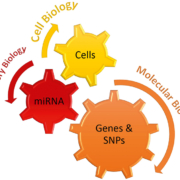
MicroRNAs (miRNAs) represent a promising source of biomarkers for GvHD because they play critical roles in the development and function of the immune system and in transplant biology (Fig. 1). MiRNAs represent a family of small (19–24 nucleotide) non-coding RNAs, which affect the regulation of gene expression in eukaryotic cells by binding to the 3´-untranslated region of target messenger RNAs [2]. They are predicted to target around 50 % of all genes and play an important role in fundamental cellular processes such as development, stem cell division, apoptosis and cancer. MiRNAs represent ideal candidates for biomarker identification in GvHD as they can be assessed using accurate and sensitive technology (e.g. NanoString/qRT-PCR), quantified in bodily fluids that require minimally invasive sample collection (e.g. serum/urine) and further investigated for biological function (e.g. target protein identification) that may expand upon our understanding of GvHD pathology. Although the field of GvHD-related miRNA research is in its infancy, recent studies have demonstrated an emerging role for miRNAs as GvHD biomarkers.
MiRNAs as biomarkers for GvHD
MiR-155 was one of the first miRNAs to be associated with the regulation of aGvHD. This miRNA is a critical regulator of inflammation, as well as adaptive and innate immune responses. In 2012, Ranganathan et al. demonstrated upregulation of miR-155 in the T-cells of mice and patients developing aGvHD following HSCT [3]. Serum expression levels also correlated with GvHD severity, and serum IFN-gamma, IL-17 and IL-9 levels, suggesting the potential of miR-155 as a biomarker for aGvHD diagnosis, and as a therapeutic target. It has since been demonstrated that miR-155 expression in both donor CD8+ T-cells and conventional CD4+ CD25− T-cells is pivotal for aGvHD pathogenesis, and drives a pro-inflammatory Th1 phenotype in donor T-cells [4].
MiR-146 is increasingly being recognized as a ‘fine-tuner’ of cell function and differentiation in both innate and the adaptive immunity. MiR-146a controls innate immune cell and T-cell responses, and directly targets two adapter proteins in the nuclear factor-kappa B (NF-κB) activation pathway; tumour necrosis factor (TNF) receptor-associated factor 6 (TRAF6) and IL-1 receptor-associated kinase 1 (IRAK1) [5]. In addition, the survival and maturation of human plasmacytoid dendritic cells that are involved in GvHD can be regulated by miR-146a. With regard to GvHD, miR-146a has been shown to be upregulated in the T-cells of nice developing aGvHD, and transplanting miR-146a–/– T-cells causes increased GvHD severity, elevated TNF serum levels and reduced survival [6]. Interestingly, Stickel et al. observed downregulation of miR-146a shortly following allo-HCT in mice (day 2), followed by upregulation in T-cells later in the aGvHD reaction (days 6 and 12), which they hypothesized may be a rescue mechanism to counteract inflammation [6]. Expression of miR-146a has since been identified to show a statistical interaction with expression of miR-155 in the peripheral blood of allo-HSCT patients before disease onset, and this interaction was predictive of aGvHD incidence, further implicating its potential as a GvHD biomarker [7].
Serum expression of miR-29a has recently been implicated as a potential biomarker for GvHD. Ranganathan et al. showed in two independent cohorts that miR-29a is significantly upregulated in allo-HSCT patients at aGvHD onset compared with non-aGvHD patients, and as early as 2 weeks before symptomatic disease onset compared to time-matched controls [8]. Further investigation into the function of miR-29a showed that it binds to and activates dendritic cells, via toll-like receptor (TLR)7 and TLR8, resulting in the activation of the NF-κB pathway and secretion of pro-inflammatory cytokines. Treatment with locked nucleic acid anti-miR-29a significantly improved survival in a mouse model of aGvHD, while retaining GvL effects [8].
In 2013 an elegant study by Xiao et al. investigated miRNA expression profiles in the plasma of patients with aGvHD, compared to patients with no aGvHD, using a qRT-PCR array to include 345 miRNAs [9]. The study identified a final signature of four miRNAs (miR-423, miR-199-3p, miR-93*, and miR-377) that significantly predicted for aGvHD at 6 weeks post-HSCT, before the onset of symptoms. Furthermore, the model was associated with disease severity and poor overall survivall [9]. Gimondi et al. have also profiled circulating miRNA expression using a qRT-PCR platform, based on samples collected 28 days post-HSCT [10]. They detected 27 miRNAs that could collectively discriminate between aGvHD and non-aGvHD. MiR-194 and miR-518f were significantly upregulated in patients who later developed aGvHD, and these miRNAs were predicted to target critical pathways implicated in aGvHD pathogenesis [10]. Our laboratory has used NanoString technology to comprehensively profile the expression of n=799 mature miRNAs in the serum of patients who had undergone HSCT, to identify miRNAs with altered expression at aGvHD diagnosis (Fig. 2) [11]. Assessment of selected miRNAs was also replicated in independent cohorts of serum samples taken at aGvHD diagnosis and before disease onset to assess their prognostic potential. Expression analysis identified 61 miRNAs that were differentially expressed at aGvHD diagnosis, and miR-146a, miR-30b-5p, miR-374-5p, miR-181a, miR-20a, and miR-15a were significantly verified in an independent cohort. MiR-146a, miR-20a, miR-18, miR-19a, miR-19b, and miR-451 were also differentially expressed 14 days post-HSCT, before the onset of symptoms, in patients who later developed aGvHD. High miR-19b expression was associated with improved overall survival, whereas high miR-20a and miR-30b-5p were associated with lower rates of non-relapse mortality and improved overall survival [11]. Collectively, these miRNA profiling studies highlight that circulating biofluid miRNAs show altered expression at aGvHD onset and have the capacity to act as independent markers for prediction, prognosis and diagnosis of GvHD.
Future directions
Despite greater recognition of the potential for miRNAs as clinically adaptable biomarkers, they have not yet reached translation to the clinic. This is predominantly because of the lack of reproducibility and independent validation to date. Indeed, owing to the high degree of variability in factors when designing and performing miRNA profiling experiments, which may be attributed to clinical (patient characteristics, sampling time points and type of body fluid analysed), technical (sample preparation, miRNA profiling platform and spectrum of miRNAs profiled) and analytical (normalization strategy) factors, progress has been slow in realizing their full potential. Despite contradictory research results on the biological basis of GvHD, low patient cohorts in single transplant centre studies, insufficient characterization of GvHD and lack of understanding and knowledge of GvHD’s impact on the immune system, miRNA biomarkers continue to show promise, but many studies are still in their infancy. Future progress relies on collaboration between research groups, focusing on standardization of the samples, protocols and technologies used, which will greatly improve the reproducibility of findings allowing for extended validation of miRNAs of interest. The ultimate aim will be to diagnose GvHD and predict outcome before the onset of clinical symptoms, allowing for earlier therapy and personalized treatments and leading to reduced mortality and morbidity outcomes.
References
1. Shlomchik WD. Graft-versus-host disease. Nat Rev Immunol 2007; 7(5): 340–352.
2. Stefan LA, Phillip DZ. Diversifying microRNA sequence and function. Nature Reviews Mol Cell Biol 2013; 14(8): 475–488.
3. Ranganathan P, Heaphy CEA, Costinean S, Stauffer N, Na C, Hamadani M, Santhanam R, Mao C, Taylor PA, et al. Regulation of acute graft-versus-host disease by microRNA-155. Blood 2012; 119(20): 4786–4797.
4. Zitzer NC, Snyder K, Meng X, Taylor PA, Efebera YA, Devine SM, Blazar BR, Garzon R, Ranganathan P. MicroRNA-155 modulates acute graft-versus-host disease by impacting T cell expansion, migration, and effector function. J Immunol 2018; 200(12): 4170–4179.
5. Taganov KD, Boldin MP, Chang KJ, Baltimore D. NF-kappaB-dependent induction of microRNA miR-146, an inhibitor targeted to signaling proteins of innate immune responses. Proc Natl Acad Sci USA 2006; 103(33): 12481–12486.
6. Stickel N, Prinz G, Pfeifer D, Hasselblatt P, Schmitt-Graeff A, Follo M, Thimme R, Finke J, Duyster J, et al. MiR-146a regulates the TRAF6/TNF-axis in donor T cells during GvHD. Blood 2014; 124(16): 2586–2595.
7. Atarod S, Ahmed MM, Lendrem C, Pearce KF, Cope W, Norden J, Wang XN, Collin M, Dickinson AM. miR-146a and miR-155 expression levels in acute graft-versus-host disease incidence. Frontiers in immunology. 2016; 7: 56.
8. Ranganathan P, Ngankeu A, Zitzer NC, Leoncini P, Yu X, Casadei L, Challagundla K, Reichenbach DK, Garman S, et al. Serum miR-29a is upregulated in acute graft-versus-host disease and activates dendritic cells through TLR binding. J Immunol 2017; 198(6):2500–2512.
9. Xiao B, Wang Y, Li W, Baker M, Guo J, Corbet K, Tsalik EL, Li QJ, Palmer SM, et al. Plasma microRNA signature as a noninvasive biomarker for acute graft-versus-host disease. Blood 2013; 122(19): 3365–33675.
10. Gimondi S, Dugo M, Vendramin A, Bermema A, Biancon G, Cavane A, Corradini P, Carniti C. Circulating miRNA panel for prediction of acute graft-versus-host disease in lymphoma patients undergoing matched unrelated hematopoietic stem cell transplantation. Exp Hematol 2016; 44(7): 624–634.e1.
11. Crossland RE, Norden J, Juric MK, Green K, Pearce KF, Lendrem C, Greinix HT, Dickinson AM. Expression of serum microRNAs is altered during acute graft-versus-host disease. Front immunol 2017; 8: 308.
The authors
Rachel E. Crossland* PhD and Anne M. Dickinson PhD
Haematological Sciences, Institute of Cellular Medicine, Newcastle University, Newcastle upon Tyne, UK
*Corresponding author
E-mail: Rachel.crossland@ncl.ac.uk
Twitter: @RECrossland


