Novel strategies for clinical coagulation diagnostics and therapy monitoring
Clinical coagulation assays are an important part of anticoagulation measurements and monitoring. Despite the rise of new promising technologies, traditional coagulation assays were largely unchanged in the last decades. Here we discuss the application of microfluidics and nanotechnology to clinical coagulation diagnostics and anticoagulation therapy monitoring.
by Dr Francesco Padovani and Prof. Martin Hegner
Introduction
Fast, accurate and reliable determination of multiple coagulation parameters is crucial for a correct diagnosis of blood coagulation disorders. The two most common coagulation assays performed regularly in hospital environments are prothrombin time (PT) and activated partial thromboplastin time (aPTT). These two assays measure the time required for the onset of fibrinogen proteolysis that is followed by the formation of a fibrin network [1]. The measurement is usually performed by increased impedance or turbidity. Upon determination of an abnormal coagulation time, further testing is required (e.g. one-stage clotting assays or chromogenic substrate assays). Despite their extreme usefulness, these assays are not factor specific and they are sensitive only if the factor activity is below 50 %. Additionally, fibrinolysis, crosslinking, clot strength or initial blood plasma viscosity (important mechanical parameters that relate to coagulation) are not measured, and finally they do not evaluate or monitor acute bleeding or thrombosis risk. These drawbacks demand for the development/standardization of novel strategies that can improve the clinical diagnosis process. Global hemostasis assays such as thromboelastography (TEG), thrombin generation, and overall hemostasis potential are promising technologies that, despite being around for decades, are not routinely used by hematologists. These assays are based on bench-top devices and require dedicated clinical laboratories and qualified personnel. Novel strategies based on microfluidics and nanotechnology may enable point-of-care testing (with potential for self-testing), self-monitoring and a great reduction in sample volume needed [2].
Anticoagulation monitoring and measurement
Accurate, reliable and frequent measurement and monitoring of anticoagulant therapies such as warfarin or heparin is vital to their effectiveness. When control is poor, patients experience more complications such as joint pain, bleeding and strokes [3]. The gold standards used for assessing the level of anticoagulation control are the percent time in therapeutic range (TTR) and international normalized ratio (INR). Both of these assays rely on standardization of the patient’s PT against an international standard. TTR is usually calculated with the method by Rosendaal that employs linear interpolation to assign an INR value to each day between successive observed INR values [4]. Therefore, patients who undergo an anticoagulation therapy have to frequently assess coagulation parameters. Systematic reviews showed that self-testing and self-management are an effective and safe intervention [5]. Self-testing devices should be of simple use, provide fast and analytically accurate results, and they should require minimal amount of sample. Ideally, they should also be portable.
Novel strategies exploiting microfluidics and nanotechnology
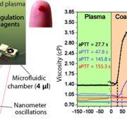
Novel approaches that employ microfluidics and nanotechnology have been developed in recent years. The main advantages of these techniques are high sensitivity and a great potential for miniaturization and point-of-care testing. Some studies proposed the use of quartz crystal microbalance (QCM) to measure the viscoelastic properties of blood plasma clot formation [6–9]. QCM consists of a quartz crystal resonator whose resonant frequency is dependent on the mass adsorbed onto the sensor and on the viscoelastic properties of the fluid surrounding the resonator. These studies showed superior performances to conventional TEG and required relatively small sample volumes. However, deconvolution of unspecific protein adsorption and liquid viscoelastic properties are very complex, hindering the potential to accurately measure clot strength development during coagulation. Other studies employed surface plasmon resonance (SPR) detection. SPR is a popular technology in the field of biomarker detection. A polarized light beam hits a glass/liquid interface causing an electromagnetic field exiting the glass. If a thin metal film is applied between the glass and the liquid surface plasmons are excited. The reflected light is collected by a sensor and upon receptor/target recognition the reflectivity curve shifts [10]. Extrapolation of viscoelastic parameters is not feasible. To the best of our knowledge, only PT time was measured using this technology [11]. Our laboratory exploited nanomechanical resonators to quantify coagulation parameters. The resonators are arrays of microcantilevers (beams clamped at one end) that oscillate at high speed. When immersed in a fluid, the viscosity and density can be measured in real time by tracking quality factor and resonant frequency of the oscillation [12]. By combining microfluidics technology, ensuring uniform mixing of coagulation reagents, with a high degree of automation and accurate extrapolation of the results, nanoresonators demonstrated great ability to measure clinically relevant coagulation parameters [13]. Along with PT and aPTT, other parameters are measured within the same test run, such as initial plasma viscosity, clot strength (final viscosity), initial and final coagulation rates. For example, patients with severe hemophilia showed a low initial plasma viscosity, low clot strength (bleeding), and low coagulation rates. By mixing hemophiliac patients’ plasma with 30 % of normal control the coagulation rates and the clot strength were improved, but not completely restored indicating the degree of severity (Fig. 1). To detect deficiencies of specific factors, an immunoassay can be integrated in situ allowing for diagnosis of factor deficiency within a single test run. Furthermore, the diagnostic array can be reused repeatably by regeneration in a cleaning solution [13]. The same microcantilever technology was applied to measure fibrinolysis in real time. It is well known that impaired function of the fibrinolytic system increases the risk of thrombosis [14]. By pre-mixing a patient’s blood plasma with tissue plasminogen activator and performing a PT (or aPTT) assay, the PT (or aPTT) and the following induced fibrinolysis can be measured. Parameters such as starting clot strength, final dissolved clot strength and 50 % lysis time (Fig. 2) provide useful information for assessing the patient’s thrombotic risk. Finally, anticoagulation treatment (heparin) was measured with low and high concentration of heparin mixed with normal control plasma (Fig. 3). Potentially, a patient under anticoagulation treatment could self-monitor their status and self-manage their therapy according to the results. For example, the final clot strength could indicate bleeding risk and the therapy can be adjusted to suit the particular needs of the specific patient (personalized medicine). All these measurements were performed with a low sample volume (<20 µl) and a high degree of automation (reducing operator intervention and complexity).
Summary
Anticoagulation measurement and monitoring employs assays that have gone largely unchanged for decades. The rise of new technologies such as microfluidics and nanotechnology carry great potential for integration with standard clinical assays. Global hemostasis assays could pave the way for an improvement in the current clinical coagulation diagnostics. Miniaturization, personalized medicine, point-of-care testing, automation, self-testing and self-monitoring are all interesting approaches that could overcome current drawbacks of gold standards in coagulation measurements. However, all these strategies require more standardization and more clinical studies to assess and exploit their potential.
Figure 1. Representation of the suspended microresonators oscillating at high speeds (approx. 300 kHz) and microfluidics set-up. Clot strength (viscosity) curves over time for normal control samples, mild hemophilia and severe hemophilia patients’ plasma during activated partial thromboplastin time (aPTT) assays performed with nanoresonators. The array of sensors is first immersed in human blood plasma (green area) and then, at time 0 s, coagulation is triggered with the specific reagents (orange area). Final clot strength, coagulation rates and aPTT values are dependent on the degree of severity. (Padovani F, Duffy J, Hegner M. Nanomechanical clinical coagulation diagnostics and monitoring of therapies. Nanoscale 2017; 9(45): 17939–17947 [13] – Reproduced by permission of The Royal Society of Chemistry.)
Figure 2. Clot strength developing over time for tissue plasminogen activator (tPA) assisted fibrinolysis. Normal control plasma was mixed with a 350 ng/ml tPA solution. After the measurement of the plasma viscosity, the coagulation is triggered at time 0 s with PT reagents. As soon as the coagulation is triggered, the clot strength increases, but at the same time the activity of tPA starts to lyse the fibrin network. After approx. 32 min, the clot is completely dissolved and the final strength is lower than the starting plasma viscosity. This difference is due to the fibrin breakage into soft fibrin particles that have no viscosity. Some of the parameters that can be extracted are PT (see zoom plot), starting clot strength (C+B), final dissolved clot strength (C), and time (50 % Ly) required to reach half-clot strength (50 % B). (Padovani F, Duffy J, Hegner M. Nanomechanical clinical coagulation diagnostics and monitoring of therapies. Nanoscale 2017; 9(45): 17939–17947 [13] – Reproduced by permission of The Royal Society of Chemistry.)
Figure 3. Effects of heparin on the clot strength development during an aPTT test. After measurement of plasma viscosity, coagulation is triggered at time 0 s with aPTT reagents. Higher concentrations of heparin cause a more prolonged aPTT but the final clot strength is always in the normal range. (Padovani F, Duffy J, Hegner M. Nanomechanical clinical coagulation diagnostics and monitoring of therapies. Nanoscale 2017; 9(45): 17939–17947 [13] – Reproduced by permission of The Royal Society of Chemistry.)
References
1. McPherson RA, Pincus MR. Henry’s clinical diagnosis and management by laboratory methods, 23rd edn (E-book). Elsevier Health Sciences 2017.
2. Al-Samkari H, Croteau SE. Shifting landscape of hemophilia therapy: implications for current clinical laboratory coagulation assays. Am J Hematol 2018; 93(8): 1082–1090.
3. Connolly S, Pogue J, Eikelboom J, Flaker G, Commerford P, Franzosi MG, Healey JS, Yusuf S; ACTIVE W Investigators. Benefit of oral anticoagulant over antiplatelet therapy in AF depends on the quality of the INR control achieved as measured by time in therapeutic range. Circulation 2008; 118: 2029–2037.
4. Razouki Z, Burgess JF Jr, Ozonoff A, Zhao S, Berlowitz D, Rose AJ. Improving anticoagulation measurement: novel warfarin composite measure. Circ Cardiovasc Qual Outcomes 2015; 8(6): 600–607.
5. Heneghan C, Ward A, Perera R, Self-Monitoring Trialist Collaboration, Bankhead C, Fuller A, Stevens R, Bradford K, Tyndel S, Alonso-Coello P, et al. Self-monitoring of oral anticoagulation: systematic review and meta-analysis of individual patient data. Lancet 2012; 379(9813): 322–334.
6. Lakshmanan RS, Efremov V, O’Donnell JS, Killard AJ. Measurement of the viscoelastic properties of blood plasma clot formation in response to tissue factor concentration-dependent activation. Anal Bioanal Chem 2016; 408(24): 6581–6588.
7. Lakshmanan RS, Efremov V, Cullen S, Byrne B, Killard AJ. Monitoring the effects of fibrinogen concentration on blood coagulation using quartz crystal microbalance (QCM) and its comparison with thromboelastography. SPIE Microtechnologies 2013, Genoble, France. Conference paper in Proc SPIE 8765, Bio-MEMS and Medical Microdevices 2013.
8. Bandey HL, Cernosek RW, Lee WE 3rd, Ondrovic LE. Blood rheological characterization using the thickness-shear mode resonator. Biosens Bioelectron 2004; 19(12): 1657–1665.
9. Hussain M. Prothrombin time (PT) for human plasma on QCM-D platform: a better alternative to ‘gold standard’. UK J Pharm Biosci 2015; 3(6): 1–8 (DOI: http://dx.doi.org/10.20510/ukjpb/3/i6/87830).
10. Hansson KM, Tengvall P, Lundström I, Rånby M, Lindahl TL. Surface plasmon resonance and free oscillation rheometry in combination: a useful approach for studies on haemostasis and interactions between whole blood and artificial surfaces. Biosens Bioelectron 2002; 17(9): 747–759.
11. Hansson KM, Vikinge TP, Rånby M, Tengvall P, Lundström I, Johansen K, Lindahl TL. Surface plasmon resonance (SPR) analysis of coagulation in whole blood with application in prothrombin time assay. Biosens Bioelectron 1999; 14(8–9): 671–682.
12. Padovani F, Duffy J, Hegner M. Microrheological coagulation assay exploiting micromechanical resonators. Anal Chem 2016; 89(1): 751–758.
13. Padovani F, Duffy J, Hegner M. Nanomechanical clinical coagulation diagnostics and monitoring of therapies. Nanoscale 2017; 9(45): 17939–17947.
14. Meltzer ME, Doggen CJ, de Groot PG, Rosendaal FR, Lisman T. The impact of the fibrinolytic system on the risk of venous and arterial thrombosis. Semin Thromb Hemost 2009; 35(05): 468–477.
The authors
Francesco Padovani PhD and Martin Hegner*PhD
Centre for Research on Adaptive Nanostructures and Nanodevices (CRANN), School of Physics, Trinity College Dublin, Dublin, Ireland
*Corresponding author
E-mail: hegnerm@tcd.ie


