Zika virus: current methods of detection and corresponding limitations
Zika virus (ZIKV) has recently become a global threat owing to the link between infection, Guillain–Barré syndrome and serious neurological defects in unborn fetus and infants. There are major challenges associated with the detection methods that are currently available for the virus, and there is no point-of-care test to accurately and quickly detect ZIKV. Herein, we describe the advantages and disadvantages of the methods that are used presently, and provide an insight into developing technologies that will yield improved detection in the future.
by Devon Pawley, Dr Emre Dikici, Dr Sapna Deo and Prof. Sylvia Daunert
Background
Infectious diseases are a serious public health concern and are the leading cause of death in low income countries [1]. The World Health Organization (WHO) declared the potential impact of the Zika virus (ZIKV) a global public health emergency in 2016, and considers the virus an ongoing threat [2]. Of particular concern is its association with Guillain–Barré syndrome and the link between ZIKV infection of pregnant women and microcephaly, neurological impairment and distress in their offspring [3, 4].
The ZIKV belongs to the genus Flavivirus, and is most commonly transmitted via different species of mosquitoes of the Aedes genus frequently found in tropical environments [5, 6]. The virus has also been shown to be transmitted from mother to fetus, as well as during sexual intercourse between individuals through bodily fluids [7]. The virus is closely related to other flaviviruses, such as the dengue virus (DENV), yellow fever virus (YFV), Japanese encephalitis virus (JEV) and West Nile virus (WNV), which often complicates correct diagnosis of ZIKV [8]. Although the virus was discovered in Uganda in 1947, the potential for the virus to infect mammals was not described until 1971 [9, 10]. Interestingly, the first clinical reports of perinatal transmission and association with Guillain–Barré syndrome due to ZIKV occurred in 2013 in French Polynesia following a major change in the virus epidemiology [11–14]. This outbreak was complicated by concurrent outbreaks of patients of DENV and chikungunya virus (CHIKV) transmitted by the same Aedes mosquito vector [15]. Since then, other reports from Brazil have chronicled a rapidly spreading epidemic that, once more, co-exists with transmission of DENV and CHIKV, and is characterized by fever, conjunctivitis, and a maculopapular rash [16]. More ominously, there are reports of microcephaly and ocular damage in aborted fetuses and infants born to mothers infected with ZIKV. In these cases, evidence of ZIKV infection came from the recovery of the virus from amniotic fluid, placental, and brain tissue. Additionally, it is known that the virus can persist in body fluids such as urine, saliva, and semen beyond the short time (<7 days) that it is present in blood, which becomes an important consideration when developing methods of ZIKV detection [17, 18].
Developing rapid diagnostics is central to prevent and control ZIKV spread, while also providing women with the necessary information to make informed decisions regarding pregnancy. It is particularly important to distinguish ZIKV infection from that of the structurally related DENV in areas where DENV is endemic and ZIKV is increasing in prevalence. Regions with the highest incidence of ZIKV infection also tend to be resource-limited. There is, therefore, an urgent and unmet need for rapid, simple, on-site, and cost-effective diagnostics that can specifically identify ZIKV and ZIKV-specific antibody (Ab) responses in body fluids.
Current ZIKV detection methods, although rapid (<30 min), are not cost effective and require specialized equipment and trained personnel. These methods are not ideal in resource-limited settings where the virus is frequently found. Additionally, these methods are regularly used concurrently for detection of ZIKV in more than one bodily fluid, most commonly urine and serum, to accurately identify the presence of the virus. Because 20–25% of infected individuals do not demonstrate symptoms, the short window of time in which ZIKV is actively present in the body is often missed [7]. Thus, tests for previous exposure to ZIKV are also performed in conjunction with tests for active infection. It is important to note that test development, validation, and optimization have proven difficult thus far due to the low amount of samples available.
Current ZIKV tests and their limitations
RNA nucleic acid tests (NATs)
The presence of active ZIKV can usually be detected early in the infection in bodily fluids using RNA NATs, such as the Trioplex real-time polymerase chain reaction (RT-PCR) Assay, loop-mediated isothermal amplification (LAMP), nucleic acid sequence-based amplification (NASBA), reverse-transcription isothermal recombinase polymerase amplification (RPA) and reverse-transcription strand-invasion based amplification (RT-SIBA) assay [19, 20]. The Trioplex RT-PCR is currently the test used by the Centers for Disease Control and Prevention (CDC) for evaluating symptomatic pregnant women in conjunction with IgM serology. Briefly, the viral RNA is first converted to cDNA via reverse transcription. If the sample contains the desired DNA sequence, a specially designed probe will bind to the target area and is detected via fluorescence. RNA nucleic acid testing is highly sensitive and can identify extremely low concentrations of viral RNA, 1.93×10⁴ genome copy equivalents per millilitre of serum according to the CDC, present during the first 10 days of ZIKV infection (21). However, NATs require expensive machinery, technical expertise, and are associated with high costs. Additionally, because viral RNA degrades rapidly in the body, NATs cannot detect prior exposure to ZIKV. Under updated recommendations of the CDC, negative NATs should be repeated with new sample extractions because of the low levels of virus present during infection.
Plaque-reduction neutralization test (PRNT)
PRNTs involve an intensely laborious process that is performed by the CDC or at a laboratory designated by CDC to detect neutralizing antibodies of a virus. If a sample has a negative ZIKV NAT and a non-negative or inconclusive serology result, a PRNT is required. PRNTs take several days to deliver a result as the process involves mixing the sample with live virus, growing this treated sample in a dish over a monolayer of host cells, and leaving the plate to incubate until plaques grow. Plaques grow when the sample added contains neutralizing antibodies, indicating previous exposure to the virus. Besides the inherent downfall of the time it takes from sample collection to plaque identification, PRNTs require specific equipment, trained personnel and do not provide information on active
ZIKV infection.
Serologic test for ZIKV
The first antibodies produced in response to initial exposure to ZIKV, IgMs, are manifested towards the end of the first week of infection. These antibodies, as well as neutralizing antibodies, can be detected via the Zika IgM Antibody Capture Enzyme-Linked Immunosorbent Assay (MAC-ELISA). A plate is coated with the anti-IgM capture, the patient’s sample is added and detection is achieved by consequential addition of an enzyme-conjugated anti-viral antibody. The enzyme interacts with a chromogenic substrate producing a colorimetric change, which can then be detected using a spectrophotometer. Important limitations to address include (1) length of assay time (2.5 days to complete); (2) detection of previous exposure to ZIKV only rather than active infection; (3) occurrence of false-negative and false-positive results. False-negatives occur when the samples were collected before IgMs have been generated, usually 4 days post-onset of symptoms or when the samples were collected after IgMs levels have fallen below detectable levels, approximately 12 weeks post-onset of symptoms. Equally, false-positives occur due to cross-reactivity with structurally similar antigens, most commonly other flaviviruses, such as DENV. Follow-up testing is necessary to rule out a false-positive result.
Active infection ELISA
Active ZIKV can be detected using a sandwich-format ELISA. Specific anti-ZIKV antibodies sandwich the virus, if it is present in the sample, and can be detected via an enzyme-conjugated secondary antibody in the same manner as the MAC-ELISA. Until recently, developing an accurate active infection ELISA proved difficult owing to the lack of specific antibodies towards ZIKV, which caused high instances of cross-reactivity with other structurally similar flaviviruses.
The previously described methods are conducted under an ‘Emergency Use Authorization’ issued by the FDA except for the active infection ELISA. In collaboration with Dr David Watkins and Dr Esper Kallas, our lab is working on developing a highly specific active infection ELISA using monoclonal antibodies isolated from ZIKV-infected patients in Sao Paulo, Brazil, that bind only to ZIKV and no other flaviviruses. Currently, our assay is under optimization to detect levels of ZIKV in urine and serum samples.
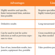
The advantages and limitations of the methods of ZIKV detection discussed above are summarized in Table 1.
Ongoing and future developments: point-of-care testing for active infection for ZIKV
Recently, paper-based detection methods have gained considerable interest because of the low cost, portability, stability at various storage conditions, and ease of use associated with their handling. These testing platforms do not require external equipment, allowing them to be carried out in remote and resource-limited areas, such as those where ZIKV flourishes. Thus, there is an emphasis on the translation of common assay principles to more portable and affordable platforms.
Lateral flow assays employ ELISA principles, and, as such, antibodies that are selective towards the desired antigen are immobilized onto a membrane. Briefly, the primary and secondary antibodies are dispensed onto the membrane via inkjet technologies and function as the test and control lines, respectively. The top portion of the membrane is laminated with an adsorbent pad to facilitate capillary action. A separate set of selective primary antibodies are conjugated to detection molecules such as gold nanoparticles, latex particles or coloured cellulose nanobeads and are immobilized onto the conjugate pad. The sample is added to the sample pad and then migrates, via capillary action, through the membrane to the conjugate pad. If the sample contains the antigen, the dried primary Ab conjugated to the coloured particles will be remobilized and the antigen will bind to these conjugated primary antibodies. The formed complexes will flow through the reaction matrix, which is usually a porous matrix such as nitrocellulose. The labelled antigen will then be captured by the immobilized primary antibodies forming a coloured band (Fig. 1). The control line will bind the coloured labelled primary antibodies regardless of the presence of antigen. This verifies that the test is working properly and the labelled conjugate can flow and bind to its respective antibody pair. When the antigen is present, the antibody/bead complex will bind to the antigen, and this Ab/antigen complex is captured by the antibody that is immobilized as the test line. One line at the control region indicates a functional but negative test and two lines indicates a functional positive test. Using our highly specific anti-ZIKV antibodies, we have additionally developed a sandwich-format lateral flow assay for the detection of ZIKV in urine that is currently under optimization.
DNA/RNA detection methods on paper are also of particular interest because of the high selectivity of hybridization. In 2015, the Whitesides group described a novel “paper machine” device that uses LAMP to detect a signal using a hand-held UV source and camera phone [22]. The paper-based device costs $1.83, an extreme improvement when compared with traditional nucleic acid testing. The only drawback of this device is that it requires incubation steps at 65 °C throughout the assay to dry the reagents present on the paper strip, which sometimes can be challenging in a point-of-care situation. Furthering the research on paper-based methods of viral RNA detection, our group described a different paper-based platform that has only one step involving incubation in a boiling water bath [23]. We have continued our pursuit to develop a point-of-care paper-based viral detection system and have constructed another test that utilizes RPA and requires incubation at much lower temperature, namely at 37 °C.
The threat of ZIKV creating serious health issues has not lessened and continues to afflict women who are pregnant and wish to become pregnant. Without proper methods of detection, the virus is difficult to characterize, document, and study. While many of the progressive paper-based platforms described herein are promising, none are currently FDA approved and on the market for use for the detection of ZIKV. It is, therefore, imperative that researchers continue to investigate and design innovative detection methods that can detect ZIKV in an easy, accurate, and affordable manner.
References
1. The top 10 causes of death. World Health Organization 2018; http: //www.who.int/mediacentre/factsheets/fs310/en/index1.html.
2. Gulland A. Zika virus is a global public health emergency, declares WHO. BMJ. 2016; 352: i657.
3. Brasil P, Pereira JP Jr, Moreira ME, Ribeiro Nogueira RM, Damasceno L, Wakimoto M, Rabello RS, Valderramos SG, Halai UA, et al. Zika virus infection in pregnant women in Rio de Janeiro. N Engl J Med 2016; 375(24): 2321–2334.
4. Štrafela P, Vizjak A, Mraz J, Mlakar J, Pižem J, Tul N, Županc TA, Popović M. Zika virus-associated micrencephaly: a thorough description of neuropathologic findings in the fetal central nervous system. Arch Pathol Lab Med 2017; 141(1): 73–81.
5. Boorman JP, Porterfield JS. A simple technique for infection of mosquitoes with viruses; transmission of Zika virus. Trans R Soc Trop Med Hyg 1956; 50(3): 238–242.
6. Haddow AJ, Williams MC, Woodall JP, Simpson DI, Goma LK. Twelve isolations of Zika virus from Aedes (Stegomyia) africanus (Theobald) taken in and above a Uganda forest. Bull World Health Organ 1964; 31: 57–69.
7. Singh RK, Dhama K, Karthik K, Tiwari R, Khandia R, Munjal A, Iqbal HMN, Malik YS, Bueno-Marí R. Advances in diagnosis, surveillance, and monitoring of Zika virus: an update. Front Microbiol 2017; 8: 2677.
8. Faye O, Freire CC, Iamarino A, Faye O, de Oliveira JV, Diallo M, Zanotto PM, Sall AA. Molecular evolution of Zika virus during its emergence in the 20(th) century. PLoS Negl Trop Dis 2014; 8(1): e2636.
9. Bell TM, Field EJ, Narang HK. Zika virus infection of the central nervous system of mice. Arch Gesamte Virusforsch 1971; 35(2): 183–193.
10. Wikan N, Smith DR. Zika virus: history of a newly emerging arbovirus. Lancet Infect Dis 2016; 16(7): e119–e126.
11. Besnard M, Lastere S, Teissier A, Cao-Lormeau V, Musso D. Evidence of perinatal transmission of Zika virus, French Polynesia, December 2013 and February 2014. Euro Surveill 2014; 19(13): pii: 20751.
12. Cao-Lormeau VM, Roche C, Teissier A, Robin E, Berry AL, Mallet HP, et al. Zika virus, French Polynesia, South Pacific, 2013. Emerg Infect Dis 2014; 20(6): 1085–1086.
13. Musso D, Nilles EJ, Cao-Lormeau VM. Rapid spread of emerging Zika virus in the Pacific area. Clin Microbiol Infect 2014; 20(10): O595–O596.
14. Oehler E, Watrin L, Larre P, Leparc-Goffart I, Lastere S, Valour F, Baudouin L, Mallet H, Musso D, Ghawche F. Zika virus infection complicated by Guillain-Barre syndrome–case report, French Polynesia, December 2013. Euro Surveill 2014; 19(9): pii: 20720.
15. Roth A, Mercier A, Lepers C, Hoy D, Duituturaga S, Benyon E, Guillaumot L, Souares Y. Concurrent outbreaks of dengue, chikungunya and Zika virus infections – an unprecedented epidemic wave of mosquito-borne viruses in the Pacific 2012-2014. Euro Surveill 2014; 19(41): pii: 20929.
16. Cardoso CW, Paploski IA, Kikuti M, Rodrigues MS, Silva MM, Campos GS, Sardi SI, Kitron U, Reis MG, Ribeiro GS. Outbreak of exanthematous illness associated with Zika, chikungunya, and dengue viruses, Salvador, Brazil. Emerg Infect Dis 2015; 21(12): 2274–2276.
17. Gourinat AC, O’Connor O, Calvez E, Goarant C, Dupont-Rouzeyrol M. Detection of Zika virus in urine. Emerg Infect Dis 2015; 21(1): 84–86.
18. Musso D, Roche C, Nhan TX, Robin E, Teissier A, Cao-Lormeau VM. Detection of Zika virus in saliva. J Clin Virol 2015; 68: 53–55.
19. Eboigbodin KE, Brummer M, Ojalehto T, Hoser M. Rapid molecular diagnostic test for Zika virus with low demands on sample preparation and instrumentation. Diagn Microbiol Infect Dis 2016; 86(4): 369–371.
20. Mauk MG, Song J, Bau HH, Liu C. Point-of-care molecular test for Zika infection. Clin Lab Int 2017; 41: 25–27.
21. Mansuy JM, Mengelle C, Pasquier C, Chapuy-Regaud S, Delobel P, Martin-Blondel G, Izopet J. Zika virus infection and prolonged viremia in whole-blood specimens. Emerg Infect Dis 2017; 23(5): 863–865.
22. Connelly JT, Rolland JP, Whitesides GM. “Paper machine” for molecular diagnostics. Anal Chem 2015; 87(15): 7595–7601.
23. Zhang DH, Broyles D, Hunt EA, Dikici E, Daunert S, Deo SK. A paper-based platform for detection of viral RNA. Analyst 2017; 142(5): 815–823.
The authors
Devon Pawley, Emre Dikici PhD, Sapna Deo PhD, Sylvia Daunert PhD
Department of Biochemistry and Molecular Biology,
Miller School of Medicine, University of Miami,
Miami, FL 33136, USA
*Corresponding author
E-mail: sdaunert@med.miami.edu


