Multiplex determination of ANA and cytoplasmic antibodies according to ICAP
by Dr Jacqueline Gosink
The international consensus on standardized nomenclature of human epithelial cell (HEp-2 cell) patterns in indirect immunofluorescence (ICAP, www.anapatterns.org) defines fifteen nuclear patterns, nine cytoplasmic patterns and five mitotic patterns which are relevant for the diagnosis of various autoimmune diseases. Furthermore, the consensus stipulates that autoantibodies detected by indirect immunofluorescence on HEp-2 cells should be confirmed by additional monospecific tests. Two new immunoblots have been developed for multiparametric autoantibody characterization. The EUROLINE ANA Profile 23 provides simultaneous confirmation of 23 autoantibodies that give rise to the nuclear patterns defined by ICAP. The EUROLINE Cytoplasm Profile allows detection of 10 autoantibodies against cytoplasmic and mitochondrial antigens, some of which are not easily recognized in the HEp-2 screening.
ICAP classification of HEp-2 patterns
Autoantibodies against cell components are important markers for the diagnosis and differentiation of systemic autoimmune diseases and other diseases. The gold standard for detection of anti-nuclear antibodies (ANA) and anti-cytoplasmic autoantibodies is the indirect immunofluorescence test (IIFT) on HEp-2 cells. Different antibodies give rise to characteristic staining patterns on the cells, depending on the cellular location and properties of the antigenic target. Analysis of primate liver tissue alongside the HEp-2 cells allows reciprocal verification and differentiation of the IIFT results and identification of unexpected additional antibodies.
In the ICAP consensus, antibodies detected on HEp-2 cells are divided into three major categories, namely nuclear, cytoplasmic and mitotic fluorescence patterns. Each category is subdivided into groups and subgroups of patterns, creating a classification tree. Each pattern and subpattern is assigned an anti-cell (AC) pattern code from AC-1 to AC-29. The pattern classification provides information about the likely target antigens and thus possible disease indications.
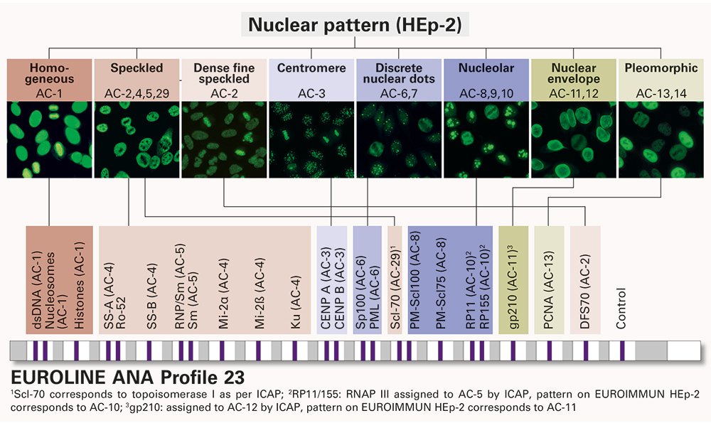
Nuclear patterns have the codes AC-1 to AC-14 plus the newly defined AC-29 and are classified into seven main groups: homogeneous, speckled, centromere, nucleolar, nuclear envelope, and pleomorphic. Autoantibodies in this category occur especially in systemic autoimmune rheumatic diseases (SARD), systemic lupus erythematosus (SLE), mixed connective tissue diseases (MCTD), Sjögren’s syndrome (SjS), systemic sclerosis (SSc), polymyositis (PM) and dermatomyositis (DM).
Cytoplasmic patterns are coded AC-15 to AC-23 and comprise five groups: fibrillar, speckled, reticular/mitochondrion-like (AMA), Polar/golgi-like, and rods and rings. Cytoplasmic antibodies are relevant in the diagnosis of SLE, PM/DM, primary biliary cirrhosis (PBC) and other diseases.
Autoantibodies against mitotic structures are assigned the codes AC-24 to AC-28. Some mitotic patterns are not exclusively associated with mitosis but exhibit very distinct features in mitotic cells. Mitotic antibodies are rare occurrences in diseases such as SSc, SLE, SjS and Raynaud’s phenomenon.
AC-0 illustrates a negative sample.
Monospecific antibody characterization
Autoantibodies can be differentiated and characterized using various monospecific tests, for example, IIFT with single purified antigen substrates or enzyme immunoassays such as ELISA or immunoblot. In IIFT, monospecific substrates (e.g. SS-A, SS-B, Scl-70, Jo-1, ribosomal P-proteins, RNP/Sm, Sm) can be analysed in parallel to the HEp-2 cells as Biochip Mosaics, thus no additional incubations are required. ELISA and immunoblot profiles provide combinations of highly defined antigens for parallel monospecific determination of different antibodies. Immunoblots are particularly well suited to this application, as they allow a huge range of different antibodies to be analysed in a single test. EUROLINE blots are fitted with individual membrane chips, thus allowing antigens with widely differing properties to be combined on one test strip. The antigen preparations and membrane fragments are individually optimized to provide maximum detection efficiency for each parameter. Thus, the profiles can be compiled according to the precise application.
ANA confirmation
The EUROLINE ANA Profile 23 (Figure 1) provides multiplex confirmation and differentiation of the most important ANA in systemic autoimmune diseases. This broad profile is the first confirmatory assay to cover all of the nuclear patterns defined in the ICAP consensus. The test antigens comprise dsDNA, nucleosomes, histones, SS-A, Ro-52, SS-B, RNP/Sm, Sm, Mi-2α, Mi-2β, Ku, CENP A, CENP B, Sp100, PML, Scl-70, PM-Scl100, PM-Scl75, RP11, RP155, gp210, PCNA and DFS70.
Cytoplasmic and mitochondrial antibody detection
The EUROLINE Cytoplasm Profile (Figure 2) allows parallel detection of autoantibodies against 10 cytoplasmic and mitochondrial antigens that give rise to some of the cytoplasmic patterns described in the ICAP consensus. The antigens comprise AMA-M2, M2-3E, ribosomal P-proteins, Jo-1, SRP, PL-7, PL-12, EJ, OJ and Ro-52. This profile is the most comprehensive assay for detection of cytoplasmic and mitochondrial autoantibodies. Since cytoplasmic antibodies are difficult to recognize in IIFT in some cases, their monospecific detection is of particular importance. The immunoblot is optimally performed in parallel to the IIFT HEp-2 screening to avoid overlooking any cytoplasmic antibodies present.
Fully automated immunoblot processing
The immunoblots for confirmation of ANA and anti-cytoplasmic antibodies can be processed manually or fully automatically, for example, using the EUROBlotOne. This device provides complete automation of all steps, from sample data entry to report release. Up to 44 strips can be incubated per run, and it is possible to combine different profiles in a single run. Preanalytical errors, due to wrong positioning of samples, are avoided thanks to the integrated barcode scanner. Moreover, the device automatically takes pictures of the strips with the integrated camera, and evaluates, interprets and archives them with the user-friendly EUROLineScan software.
Conclusions
The ICAP statement is an on-going process which will be adapted in the future based on feedback from clinicians. The long-term goal is to develop it into a global standard for the reporting of autoantibodies on HEp-2 cells. The EUROLINE system is highly suited for monospecific differentiation and characterization of autoantibodies, offering a multiplex format, ease of use and automatability. As the classification system matures, the profiles can be easily adapted, for example by supplementing them with important new antigens. Notably, the EUROLINE is the most frequently used ANA confirmatory test in various national quality assessment schemes, reflecting its exceptional quality and successful application in clinical diagnostic laboratories.
The author
Jacqueline Gosink, PhD
EUROIMMUN AG, Seekmap 31,
23560 Luebeck, Germany
www.euroimmun.com


