Evaluation of UriSed mini semi-automated urine microscopy analyser
Urinalysis may provide evidence of significant renal disease in asymptomatic patients. The microscopic urinalysis is vital to making diagnoses in many asymptomatic cases, including urinary tract infection, urinary tract tumours, occult glomerulonephritis, and interstitial nephritis.
Presence or absence of different particles in urine sediment is crucial for clinical decision making. Urine sediment cells or particles provide important information for the diagnosis of renal or urinary diseases [1]. UriSed Technology is a unique solution in the market for the automation of sediment analysis, making traditional manual microscopy automatic [2]. The UriSed analysers provide a reliable and reproducible solution since 2007 [3]. As a new category instrument based on the improved UriSed Technology, the semi-automated UriSed mini was introduced in the market in 2015. In the present study, we evaluated the analytical performance of UriSed mini Semi-Automated Urine Microscopy Analyser (Manufactured by 77 Elektronika Kft., Budapest) and compared the results to those from manual microscopy using standardized KOVA counting chambers.
UriSed mini provides quantitative Red Blood Cell (RBC) and White Blood Cell (WBC) results, and semi-quantitative results for all other particle types: Squamous Epithelial Cells (EPI), Non-squamous Epithelial Cells (renal tubular and urothelium cells) (NEC), Crystals (CRY): Calcium oxalate dihydrate (CaOxd), Calcium oxalate monohydrate (CaOxm), Uric acid (URI), and Triple-phosphate crystals (TRI), Hyaline casts (HYA), Pathological casts (PAT), Bacteria (cocci-like and rod-like) (BACc, BACr), Yeasts (YEA), Spermatozoa (SPRM) and Mucus (MUC) [4].
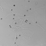
The instrument throughput is up to 60 samples per hour. Preparation of the UriSed mini analyser for measurement takes only some minutes, the only consumables are the patented disposable cuvettes for sample investigation and a manual pipette with appropriate pipette tip. 175 µl of urine sample is dispensed manually into the cuvette, further steps of the measurement sequence are completely automatic: spinning the cuvette for a few seconds gently deposits formed elements into a monolayer at the bottom of the cuvette. The built-in digital camera takes 15 independent images at different positions of the sediment layer. These whole viewfield images are evaluated by a neural-network based image processing software.
Material and methods
Analysis of 311 samples was performed to evaluate UriSed mini analytical performance compared to the manual microscopy urine examination method. Both measurements were carried out with the same anonymous urine samples. Fresh, native urine samples were used, that were typically held for no more than 4 hours before being analysed, as recommended in the relevant guidelines [5,6] to prevent change in the morphology of the particles. Samples were mixed until homogeneous and then split and run on each measuring procedure as close to the same time as possible. The standardized microscopic urinalysis of native samples (Level 3) was followed by using a KOVA counting chamber. The particle concentration for all particle types was evenly distributed in the evaluated urine samples. Carry-over, precision, diagnostic tests such as sensitivity, specificity, diagnostic accuracy, concordance and one category concordance were investigated according to well-established protocols [7].
Results
No carry-over was detected in any of the samples due to the single-use disposables. UriSed mini has better precision than microscopy at all of the tested RBC and WBC concentrations. The majority of all coefficients of variation obtained for within-series imprecision (CV) using UriSed mini was 4-24% while 5,5-67% in the case of manual microscopy [8]. Good correlation can be found between UriSed mini and manual counting chamber for formed elements. The Pearson correlation of quantitative parameters are 0.97 (RBC), 0.95 (WBC). The clinical evaluation of UriSed mini was based on McNemar test and concordance study. The results are shown in the following table.
Conclusion
The new UriSed mini utilizes the traditional gold standard method while eliminating the most time-consuming and operator-dependent procedures in laboratories performing manual microscopy. The semi-automated measurement process is reproducible and operator-independent. The UriSed mini semi-automated microscopy analyser requires manual sample pipetting, which makes the instrument small and simple to use. UriSed mini is a highly effective tool in a wide range of medical and clinical settings such as hospitals, clinics, accident and emergency departments and outpatient laboratories. In addition it can also serve as a backup instrument of automated sediment analysers.
References
1. Fogazzi GB. The Urinary Sediment an Integrated View Third Edition. Milano: Elsevier, 2010.
2. Barta Z., Kránicz T., Bayer G. UriSed Technology – A Standardised Automatic Method of Urine Sediment Analysis. European Infectious Disease 2011;5:139–42.
3. Zaman Z, Fogazzi GB, Garigali G, Croci MD, Bayer G, Kránicz T. Urine sediment analysis: analytical and diagnostic performances of sediMAX – a new automated microscopy image-based urine sediment analyser. Clin Chim Acta 2010; 411: 147-154.
4. Fogazzi GB, Garigali G. The Urinary Sediment by UriSed Technology. A New Approach to Urinary Sediment Examination. Milano: Elsevier, 2013.
5. Kouri T, Fogazzi G, Hallander H, Hofmann W, Guder WG, editors. European Urinalysis Guidelines. Scand J Clin Lab Invest 2000; 60 (Suppl 231): 1-96.
6. Clinical and Laboratory Standard Institute (ex NCCLS). Document GP16-A3 – Urinalysis; Approved guideline, 3rd ed. Wayne, PA: CLSI, 2009.
7. T. Kouri, A. Gyory, R.M. Rowan. ISLH Recommended Reference Procedure for the enumeration of Particles in Urine. Laboratory Hematology 9:58-63, 2003.
8. Haber MH, Galagan K, Blomberg D, Glassy EF, Ward PCJ, editors. Color Atlas of Urinary Sediment; An Illustrated Field Guide Based on Proficiency Testing. Chicago: CAP Press, 2010.
More information on UriSed mini is available from the manufacturer:
77 Elektronika Kft., Budapest, HUNGARY.
Email: sales@e77.hu, web: www.e77.hu
The author
Erzsébet Nagy MD,
Honorary Associate Professor
Head Physician of Central Laboratory;
Hospitaller Brothers of St. John of God
Hospital, Budapest


