


Bence Jones Proteinuria Testing: An IFCC-Based Alternative Using Quantitative Urinary Free Light Chains
Why Bence Jones Proteinuria Testing Remains Challenging
Bence Jones proteinuria (BJP), defined by urinary excretion of monoclonal immunoglobulin free light chains (ur-FLC), remains relevant in plasma cell dyscrasias and other monoclonal gammopathies. Urine immunofixation electrophoresis (ur-IFX) is essential to confirm monoclonality and identify the light-chain type, but traditional workflows may require urine concentration, manual processing and specialist interpretation. Densitometric quantification can also be limited when monoclonal bands are weak, poorly resolved or affected by protein co-migration.
The IFCC Committee on Plasma Proteins proposed an alternative approach based on immunochemical quantification of ur-FLC as an initial screening step, reserving immunofixation for samples requiring confirmation.[1] Modern nephelometric and turbidimetric assays make this strategy increasingly applicable to routine laboratory workflows.
Quantitative ur-FLC and Confirmed BJP: Complementary Concepts
Free κ and λ light chains are normally filtered by the glomerulus and largely reabsorbed in the proximal tubule. In monoclonal plasma cell disorders, production may exceed tubular reabsorptive capacity, leading to urinary excretion. However, ur-FLC concentrations are influenced not only by production, but also by renal function and urinary protein handling.[1]
Quantitative ur-FLC concentration and confirmed BJP should therefore remain distinct concepts: immunochemical assays provide numerical κ and λ values, whereas immunofixation establishes whether the pattern is monoclonal, oligoclonal or polyclonal.

An IFCC-Based Three-Step Workflow
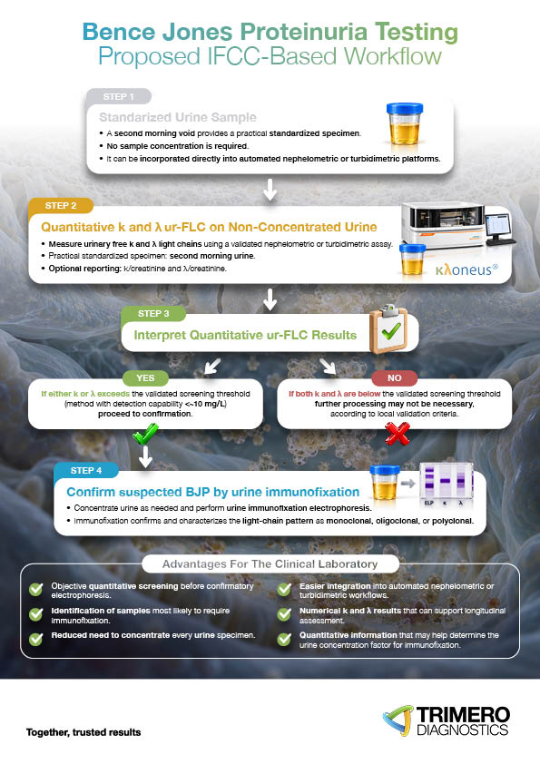
The IFCC-based pathway can be structured in three steps.
First, use a standardized specimen, preferably a second morning urine sample, and express results relative to urinary creatinine to reduce the effect of variable urine concentration.[1,2]
Second, apply a quantitative ur-FLC method with a detection capability below approximately 10 mg/L and adequate control of antigen excess.[1] If either κ or λ exceeds the validated screening threshold, the sample should proceed to confirmatory testing; if both remain below the threshold, further processing may not be necessary, depending on assay performance and laboratory validation.
Third, confirm suspected BJP by urine immunofixation.
From Quantitative Screening to Targeted Confirmation
This approach can improve workflow efficiency by directing electrophoresis and immunofixation toward the samples most likely to benefit from them. Potential advantages include objective quantitative screening, fewer samples requiring concentration, easier integration into automated platforms, numerical κ and λ results for longitudinal assessment, and more informed selection of the urine concentration factor for immunofixation.
Quantitative ur-FLC and densitometric BJP are nevertheless complementary, not interchangeable. Recent studies show that performance varies with decision limits, renal impairment, total urinary protein and low-level BJP.[2,3]
The goal of quantitative ur-FLC testing is therefore not to reproduce a densitometric BJP value, but to provide sensitive, objective information that supports screening and laboratory decision-making.
Integrating KLoneus® into the Laboratory Workflow
KLoneus® Free Light Chains assays can facilitate this workflow by providing quantitative κ and λ measurements in serum and urine on nephelometric and turbidimetric systems.
Within the IFCC-based pathway, KLoneus® can support a straightforward sequence:
Non-concentrated urine → quantitative ur-FLC → screening decision → confirmatory immunofixation when indicated.
The value of this strategy lies not in replacing immunofixation, but in combining automated quantitative screening with targeted qualitative confirmation.
With appropriate analytical validation and assay-specific decision criteria, ur-FLC testing can support a more objective, selective and structured approach to Bence Jones proteinuria assessment.
To learn more about KLoneus® Free Light Chains and explore Trimero Diagnostics’ solutions for specialized protein testing, visit www.3diag.com.
References
- Graziani M, Merlini G, Petrini C; IFCC Committee on Plasma Proteins and SIBioC Study Group on Proteins. Guidelines for the Analysis of Bence Jones Protein. Clin Chem Lab Med. 2003;41(3):338–346. doi:10.1515/CCLM.2003.054.
- Tillio PA, Lotfolahzadeh Y, Kroumova V, et al. Improving diagnostic strategies for plasma cell dyscrasias: a laboratory perspective on Bence Jones proteinuria. Clin Chim Acta. 2026;593:121310. doi:10.1016/j.cca.2026.121310.
- Michetti L, Ravasio R, Marozzi R, et al. Immunochemical measurement of urinary free light chains and Bence Jones proteinuria. Clin Chem Lab Med. 2025;63(10):2031–2042. doi:10.1515/cclm-2025-0265.