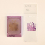
Ensuring sectioning quality in TMA analysis for the Human Protein Atlas project
The Human Protein Atlas Project is carrying out the systematic exploration of the human proteome using antibody-based proteomics, thus providing an invaluable publicly available HPA portal tool for pathology-based biomedical research. As part of the project, the Uppsala-based Science for Life Laboratory tissue profiling group has so far cut more than 200,000 slides from over 1400 tissue microarrays (TMAs). This article describes how the tissue microarrays and slides are made, and how a rotary microtome with different cutting modes and an automated Section Transfer System together ensure that high-quality, reproducible sections are generated.
by Ing-Marie Olsson, Catherine Davidson and Dr Caroline Kampf
A publicly available protein dictionary
Molecular tools developed in the research arena are making a significant contribution in the evolution of tissue-based diagnostics. Immunohistochemistry (IHC) is now well recognised as a means of enhancing morphological analysis, with protein expression patterns considered as effective diagnostic and prognostic indicators for various cancers. For example, within diagnostic pathology, IHC could determine the origin of poorly differentiated tumours and also be used to stratify tumours for optimum treatment regimes.
Consequently, the Human Protein Atlas (HPA) project was initiated in 2003 by the Knut and Alice Wallenberg Foundation to enable the systematic exploration of the human proteome using antibody-based proteomics. Since then, the publicly available HPA portal (www.proteinatlas.org) has amassed a database of millions of high resolution images showing the spatial distribution of proteins in 46 different normal human tissues and 20 different cancer cell types, as well as 47 different human cell lines. As such, the HPA can provide an invaluable tool for pathology-based biomedical research, including protein science and biomarker discovery for disease identification [1].
Tissue profiling
One of the key sites involved in this immense project is the Uppsala-based Science for Life Laboratory (SciLifeLab Uppsala) tissue profiling group [2]. This highly experienced group is focused on histopathology, with special emphasis on tissue microarray (TMA) production, immunohistochemistry and slide scanning. The enormity of profiling the human proteome requires the use of high throughput techniques, prompting the SciLifeLab team to adopt a TMA format to enable them to perform simultaneous multiplex histological analyses.
TMAs are paraffin blocks containing cores of selected tissues or cell preparations assembled together for subsequent sectioning to enable the effective and efficient utilisation of valuable tissue samples, as well as reducing the use of expensive IHC reagents. Multi-tissue blocks were first introduced by Battifora in 1986 with his ‘multitumour (sausage) tissue block’ [3]. Then in 1998, Kononen and collaborators standardised the technology and developed instrumentation which uses a sampling approach to produce tissues of regular size and shape that can be more densely and precisely arrayed [4].
As part of the HPA project the SciLifeLab Uppsala tissue profiling facility has constructed over 1400 TMAs containing over 100,000 tissue cores, in addition to 180 cellular microarrays (CMA) containing over 23,800 cell cores. Over 200,000 slides cut from these arrays have then been stained using immunohistochemical techniques, of which more than 100,000 have been scanned for further analysis. The SciLifeLab team evidently holds a great deal of practical experience in TMA production and, in fact, now offers an external TMA production service [2]. Consequently, its experts handle many different types and combinations of tissues, for which they observe that high quality sectioning is fundamental to TMA production, the primary aim of which is to amplify a scarce resource.
TMA production
The most efficient method of constructing tissue microarrays is by extracting cylinders of donor tissue with a sharp punch and then assembling them into a recipient block that has uniformly sized holes in a grid pattern. Tissue and cell microarrays are made according to a preset standard within the HPA, where paraffin blocks are used in a matrix containing from 72 up to 120 tissue cores. The standard diameter of each core is 1 mm (tissues) and 0.6 mm (cells), with a length of 2-4mm. This is achieved by using a needle to remove relevant tissue from a donor paraffin block which is then inserted into a recipient paraffin block.
Once all tissue cores are in position within the array, it is then ‘baked’ at 42ºC to melt them together into a homogenous paraffin block. This 40 minute baking period ensures that every core is merged with the melted paraffin in the block and, therefore, totally secured for sectioning into 4 µm sections prior to mounting onto glass slides. Thereafter, these multiplex tissue sections are ready for further histological analysis and final slide scanning to transf orm stained glass slides into digital high-resolution images.
Quality sectioning
When sectioning TMAs, the greatest risk of valuable tissue loss or damage can occur during transfer to a water bath. For this reason, the SciLifeLab tissue profiling group uses microtomes with a ‘waterfall’ system (Thermo Scientific HM355S and Thermo Scientific Section Transfer System) to eliminate such risks. A ‘waterfall’ automated Section Transfer System stretches sample ribbons as they are cut, whilst simultaneously transporting them from the blade into the attached circulating laminar flow bath. From this water bath, sections can be extracted and mounted onto a glass slide. Mounting two microarray sections per slide can further reduce IHC reagent usage and enhance workflow within the tissue profiling group.
By using the Section Transfer System the group routinely obtains over 200 quality sections per TMA, depending on the size of donor block and representative tissue within it. Although it is possible to obtain many more sections, for quality assurance (QA) purposes the SciLifeLab team performs a QA after every 50th section, introducing replacement cores where required to ensure that at least 85% of the tissue cores are always present.
The actual composition of a tissue array can also cause complications when sectioning, dependent on whether tissues are homogenous cancer types, or normal tissues where heterogeneity is greater. Furthermore, fatty tissue such as that from brain and breast should not remain within a warm water bath for an extended period due to risk of tissue melting. Conversely, other tissue such as skin and thyroid gland, needs to remain in the water bath for longer in order to ensure that it is sufficiently stretched.
To overcome such issues with tissue composition, SciLifeLab experts group tissues into those with similar texture and hardness when sectioning to make set up easier and improve workflow. For example, the HM355S microtome offers a choice of four mechanised cutting modes that give SciLifeLab greater control over section generation according to varying requirements. Mechanised cutting delivers the slow, smooth, even and controlled action necessary for sectioning harder consistency specimens.
A further sectioning consideration at SciLifeLab Uppsala is the fact that the TMAs are paraffin embedded. Consequently, a peltier-cooled attachment (Thermo Scientific Cool Cut) is used on the group’s microtomes to prolong the cutting period by maintaining a cool block temperature. By using such a cooling tool, 50 TMA sections can be cut consecutively in 50 minutes without the need to remove and re-cool the block on ice, again ensuring effective throughput and efficient laboratory operation.
SciLifeLab tissue profiling services
Tissue Microarrays (TMAs) are coming to the fore as an ideal means of providing multiplex tissue analysis, not only for research based applications, but also for clinical applications: identifying biomarkers for identification of disease, histological grading and detecting disease recurrence [5,6,7]. Some hospital laboratories are also starting to utilise TMAs as controls for diagnostic comparisons.
With over 100 personnel working on the HPA project alone, the SciLifeLab facility provides access to its extensive protein profiling results to laboratories throughout Sweden and beyond. In addition, leveraging their expertise gained in constructing tissue arrays for high throughput protein screening, the SciLifeLab team in Uppsala has also recently extended its capabilities to offer an external TMA production, sectioning and scanning service [2].
Working to a user specified template, the facility can turnaround 120 core duplicate arrays within 24 hours from receipt of the donor tissue blocks. The venture operates as cost neutral, utilising the team’s experience in generating high quality sections at a resolution of 2µm-10µm to provide consistent and reproducible material for downstream analysis. Since its inception, the TMA service has produced more than 100 custom arrays, supporting investigation of clinical models for a wide range of disease states, including cancer, diabetes, heart disease and neurodegenerative disorders.
Establishments utilising the tissue profiling group’s TMA services include university research, hospital and even veterinary laboratories. Such is the experience of this SciLifeLab group, it has been able to produce TMAs on almost any kind of tissue. Although bone and skin can prove difficult, the team can even produce TMAs for these by careful orientation of skin samples and decalcification of bone prior to final preparation.
Advanced technical know-how and state-of-the-art equipment, combined with a broad scientific knowledge, all mean that the SciLifeLab tissue profiling facility is ideally placed to meet high throughput, high quality TMA production needs for the HPA, whilst simultaneously ensuring service excellence for external customers.
References
1. Pontén F et al. The Human Protein Atlas – a tool for pathology. J Pathol 2008; 216(4): 387-93.
2. http://scilifelab.uu.se/technologyplatforms/Proteomic/Tissue_Profiling_Center/?languageId=1
3. Battifora H. The multitumor (sausage) tissue block: novel method for immunohistochemical antibody testing. Lab Invest 1986; 55(2): 244-8.
4. Kononen J et al. Tissue microarrays for high-throughput molecular profiling of tumor specimens. Nature Medicine 2008; 4: 844-847.
5. Rimm D et al. Cancer and Leukemia Group B Pathology Committee Guidelines for tissue microarray construction representing multicentre prospective clinical trial issues. J Clinical Oncology 2011; 29 (16): 2282-2290.
6. Schmidt L et al. Tissue microarrays are reliable tools for the clinicopathological characterisation of lung cancer tissue. Anticancer Research 2009; 29: 201-210.
7. Smith V et al. Tissue microarrays of human xenografts. Cancer Genomics & Proteomics 2008; 5: 263-274.
The authors
Ing-Marie Olsson, Team Leader
TMA production, sectioning and scanning, Human Protein Atlas (HPA), Tissue Profiling Centre, Science for Life Laboratory, Department of Immunology, Genetics and Pathology, Uppsala, Sweden
Tel. +46 18 471 5040
e-mail: ingmarie.olsson@igp.uu.se
Catherine Davidson, Sectioning Product Manager
Thermo Fisher Scientific, Anatomical Pathology, Runcorn, UK
Tel. +44 (0) 1928 534122
e-mail: catherine.davidson@thermofisher.com
www.thermoscientific.com/pathology
Dr. Caroline Kampf, Site Director Human Protein Atlas (HPA), Tissue Profiling Centre, Science for Life Laboratory, Department of Immunology, Genetics and Pathology, Uppsala, Sweden
Tel. +46 18 471 4879
e-mail: Caroline.Kampf@igp.uu.se


