Insight into advances in diagnosis of Barrett’s esophagus
Barrett’s esophagus has been established as a pre-cancerous condition that increases an individual’s risk of developing esophageal cancer, and so regular monitoring people with Barrett’s esophagus can help improve the survival rates of those who develop cancer. However, diagnosis is usually by endoscopy, which is invasive, unpleasant and resource-intensive. CLI caught up with Charlene Tang (Commercial Manager of Cyted) to discuss their new method for diagnosing and monitoring Barrett’s esophagus, which is much easier to administer and can help prioritise the use of endoscopy for patients who need it most.
What is Barrett’s esophagus and why is diagnosis important?
Barrett’s esophagus is a condition in which the cells lining the esophagus change, which is thought to be as a result of long-term exposure to acid reflux. Barrett’s esophagus is a well-defined precursor of the most common type of esophageal cancer, esophageal adenocarcinoma, and its prevalence is increasing. Around 3–13% of people with Barrett’s esophagus develop esophageal cancer – 11 times more than the average person.
Barrett’s esophagus often goes undiagnosed as its main symptoms include heartburn and indigestion. However, early-stage cancer diagnosis can dramatically improve a patient’s quality and length of life. For cancer of the esophagus, over 8 in 10 patients survive beyond 5 years if it is diagnosed and treated at an early stage, compared to just 2 in 10 patients surviving beyond 5 years if it is diagnosed at a late stage.
How is diagnosis for Barrett’s esophagus usually done?
The standard-of-care diagnosis of Barrett’s esophagus currently requires endoscopy, which is expensive, invasive and unpleasant for the patient. Endoscopies are needed for patients at higher risk of esophageal cancer and for example, factors that increase the risk include:
• family history of esophageal cancer;
• obesity;
• smoking;
• being on repeat prescription for proton-pump inhibitors;
• long-term over-the-counter use of antacids; and
• worsening or persistent heartburn symptoms.
Endoscopies are also needed for patients already diagnosed with Barrett’s esophagus who have regular surveillance to monitor the severity of their condition and for any signs of cancer. The COVID-19 pandemic paused several key healthcare services, including endoscopy, resulting in an extensive backlog of patients. In September 2021, it was reported that over 60 000 patients were on the waiting list for an upper gastrointestinal endoscopy in England. In some cases, patients are waiting several months for their endoscopy appointment. For a patient who actually has an undiagnosed cancer, the longer the wait, the more likely the cancer is to advance and worsen, and the less likely treatment will be successful. To ensure that patients at risk of esophageal cancer are found sooner and can start life-changing treatment when it is more likely to be successful, there is a clear need for an enhanced form of less-invasive and less-resource-intensive diagnostic testing.
Is there a way that this can be improved?
The Cytosponge™ (Medtronic) is a novel, inexpensive, minimally invasive diagnostic test for Barrett’s oesophagus that could revolutionize the clinical care pathway for reflux disease as an alternative to endoscopy. The test identifies the presence of trefoil factor 3 (TFF3) and tumour suppressor protein 53 (p53), which are biomarkers for precancerous changes (intestinal metaplasia and atypia/dysplasia) that indicate Barrett’s esophagus and early esophageal cancer.
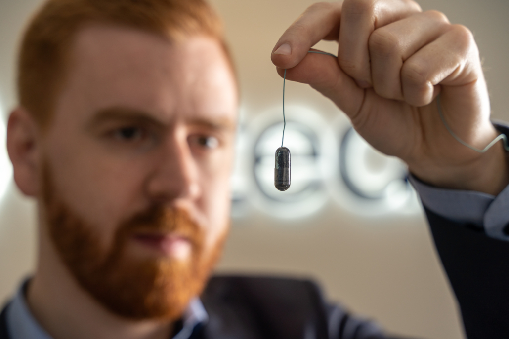
The Cytosponge is a ‘sponge-on-a-string’ device to collect cells from the esophagus. The patient swallows the pill-sized capsule, which is attached to a thread. The capsule dissolves in the stomach and releases the sponge that is attached to the thread. The mesh sphere is then pulled back up from the stomach and collects cells from the surface of the esophagus on its way. The cells are then sent to the Cyted laboratory for digital diagnostic processing and analysis using artificial intelligence (AI). The Cyto-sponge technology has been developed and supported by scientists at the University of Cambridge from conception, through a series of clinical studies to the ongoing implementation pilot with the spin-out company Cyted. The Cytosponge test has been shown to be safe and acceptable to patients in studies with a total of over 4000 patients across the UK, USA and Australia. Since clinics launched in August 2020, over 5000 patients have been tested through NHS England and NHS Scotland.
By systematically using positive Cytosponge tests to identify those who are at most risk of Barrett’s esophagus or early esophageal cancer, those patients with greatest need can be prioritized for endoscopic confirmation and subsequent treatment, supporting pathologists to deliver greater impact, more effectively.
Owing to its cost-effectiveness compared to endoscopy, this pathway is likely to result in an economic benefit to the NHS as well as to improve the patient experience and save lives through the earlier and faster detection of cancer. Use of the Cytosponge will help to relieve strain on the Barrett’s esophagus surveillance and screening/referral pathways and to recover from the increased numbers of patients endoscopy service waiting lists.
The Cytosponge test overcomes the limitations in accessibility of endoscopy. Endoscopy requires the patient to be referred to a specialist gastroenterology team in secondary care to undergo an invasive procedure that requires several hours spent in preparation and recovery, as well as introduces risk to elderly patients. In contrast, the Cytosponge procedure can be carried out by a trained nurse in an office setting and requires 10 minutes to explain and administer. Additionally, a Cytosponge test can be delivered through mobile diagnostic units (as being piloted through the Innovate UK-funded Project DELTA) and mobile practice or community nurses. Altogether, this means that the Cytosponge test can be accessed by difficult-to-reach patient populations in terms of geographical location and socio-economic factors.
How can automated intelligence help with the workload?
The analysis of the patient samples, looking for signs of disease, is comparable to finding a needle in a haystack. Even for an experienced pathologist, the process is resource-intensive and mentally rigorous.
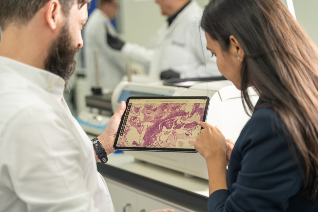
Transforming this way of working, Cyted has developed a novel, machine-learning method that will assist with sample analysis and speed up review times. First, the samples are digitized, and then the images are analysed using the digital platform. Like GPS, the machine-learning method directs pathologists to the areas of the sample that need their attention most, ensuring they focus their time and attention on the most difficult cases so they can detect disease earlier and save lives faster.
Over time, as more and more samples are analysed, the model will evolve and become even more accurate in its diagnostic results. This will reduce the number of samples that require manual analysis by a pathologist, streamlining the process, increasing the capacity of laboratories, and ultimately helping to save more lives.
Is it time for a screening programme for Barrett’s esophagus and how would screening be targeted appropriately?
The surveillance of Barrett’s esophagus has been shown to correlate with earlier stage detection of and improved survival from cancer – which is incredible. Long-standing cancer screening programmes have demonstrated their benefit, with breast cancer screening saving about 1300 lives each year in the UK. Esophageal cancer currently claims around 7990 lives per year in the UK, so the potential to save lives is huge.
Surveillance with the Cytosponge test would transform outcomes for patients, through the earlier and faster detection of a potentially lethal type of cancer and removing the need for endoscopy, an unpleasant and invasive procedure, for many patients. By contrast, screening with the Cytosponge would be a fast, minimally invasive procedure that can be carried out by trained nurses in an office setting. Therefore, there is the potential for screening to be carried out in community settings to maximize its reach, such as within the NHS Community Diagnostic Centres that are being set up across England.
By detecting cases of Barrett’s esophagus, and esophageal cancer, that would otherwise go unnoticed until they are much harder to treat, thousands of lives could be saved. The Barrett’s Esophagus Trial 3 (BEST3) clinical trial showed that offering the Cytosponge test in primary care could detect 10 times more patients with Barrett’s esophagus than standard-of-care with endoscopy.
In addition to the significant potential to transform outcomes for patients, a screening programme would reduce pressures on health services, through cost and time savings.
There is potential to reach and test a significant proportion of the population; however, to maximize outcomes a screening programme should first target those most at risk (as identified by the risk factors listed above), and then be expanded to the general public. Depending on the result of the Cytosponge test, patients may be sent for an endoscopy as urgent, with only a two-week wait.
What do you envisage for the future development of AI in pathology/digital imaging?
The power of AI and digital platforms offers truly unlimited potential to transform diagnostic pathways. Pathologists will be empowered to do more – more accurately – so they can detect disease earlier and save lives faster. Improving access to digital diagnostic services will also transform healthcare, as health systems will be able to benefit from these cutting-edge tools without facing the burden of high investment themselves. As a result, patients will receive their diagnosis much faster, and be supported to start potentially life-saving treatment earlier.
As the use and development of the deep-learning tool used in analysing Cytosponge samples continues to evolve, it will become even more accurate in categorizing samples into their level of risk, reducing workload pressures on our pathologists. The technology also isn’t limited to this one condition and, in the future, it could potentially be applied to other conditions including pancreatic, gastric and bowel cancer.
Advances in AI and digital pathology enable pathologists to be supported to push the boundaries of what is possible. As we continue to grow our pipeline across new disease areas and enhance our AI technology, digital pathology will become increasingly imbedded into diagnostic workflows in the UK and beyond, enabling all patients to benefit from the earlier and faster detection of disease.
Dr Marcel Gehrung, co-founder and CEO of Cyted, with the Cytosponge test, a ‘sponge on a string’ device Credit: Cyted
Digital image of a Cytosponge sample section featuring Alec Hirst, Cyted’s Senior Director of Business Technology, and Neha Goel, Cyted’s COO Credit: Cyted
Charlene Tang, BA, MSci, Commercial Manager Cyted
The interviewee
Charlene Tang, BA, MSci, Commercial Manager
Cyted, Cambridge, Cambridgeshire, CB1 2JH, UK
E-mail: charlene.tang@cyted.ai
For further information visit Cyted (https://cyted.ai/)


