Molecular allergology offers new opportunities – for the lab and the clinician
Molecular allergology enables quantification of IgE antibodies to single allergen protein components at the molecular level. This helps the clinician establish the cause of allergic sensitisation, evaluate the risk for severe allergic reactions and improve patient management. New tests and technologies enable the laboratory to assist in an efficient manner.
by Dr Magnus Borres
For quite some time there has been scientific interest in the individual proteins contained in an allergy source, e.g. a pollen, foodstuff or animal fur. One of the first food allergen components, Gad c 1 from cod, was purified as early as the late sixties [1]. The term Component Resolved Diagnostics was introduced in 1999 [2]. Further developments, such as the production of recombinant allergen components and the use of microarray technology, have resulted in novel practical tools for the clinician and the laboratory. The term molecular allergology is now used to describe this new breakthrough science.
There is currently great interest in this area since several research studies have shown that molecular allergy diagnostics can result in:
- a more precise diagnosis of the allergic sensitisation by identification of specific versus cross-reactive components
- improved risk assessment by identification of components linked to a high- versus low risk of inducing severe, systemic reaction; and
- changed indications for immunotherapy or clinical management.
Allergen sources are complex
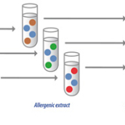
All allergen sources contain a number of different proteins, some of which can cause allergy. Each allergen component usually has several epitopes, the actual three-dimensional binding site for the corresponding antibody. Some allergen components are unique markers for a specific allergen source. Others have protein structures common to widely different species. In such cases IgE-sensitisation may be due to cross-reactivity with proteins with similar epitopes [Figure 1].
A positive extract-based IgE test does not give any information about which of the many components in the extract triggered the IgE response. Such information is of great value both for the clinician and the patient – is it a component that is present in many different allergen sources (cross-reactive sensitisation), or is it a species specific (primary) sensitisation? Furthermore, some components are prone to induce severe reactions while others cause sensitisation without clinical reactions. The stability of the component to heating and digestion also varies, and which is of great significance in food allergy.
Thanks to molecular allergology there are now good tools available for quantifying IgE sensitisation to allergen components, both tests for single components and microarray-based tests giving answers to 100+ components from a small amount of blood sample.
Single component tests
Allergen components are given designations based on the Latin family name of the species, e.g. Ara h 1 stands for the first allergen component of peanut, Arachis hypogea.
Tests for IgE antibodies to single allergen components are based on proteins purified from their natural source, or produced via recombinant techniques. Such single component tests provide high-quality, accurate, quantitative results and are therefore an essential tool for the allergy physician. They are especially useful for investigating allergy to one or a few suspected defined sources. The most comprehensive range of single component tests, ImmunoCAP Allergen Components (ThermoFisher Scientific, formerly Phadia AB, Uppsala, Sweden) contains over 90 allergen components.
Biochip technology
Microarray or biochip technology makes it possible to simultaneously assay a large number of allergen components in a minute amount of patient sample. ImmunoCAP ISAC (ThermoFisher Scientific, formerly Phadia AB, Uppsala, Sweden) is a miniaturised immunoassay platform where allergen components are immobilised in a microarray. This enables simultaneous measurements of IgE antibodies to a fixed panel of 112 components from 51 allergen sources, using only 30 µl of serum or plasma. The allergen components are spotted in triplets and covalently immobilised to a polymer coated slide. Each slide contains four microarrays, giving results for four different samples per slide [Figure 2].
The assay consists of two steps. In the first, IgE antibodies from the patient sample bind to the immobilised allergen components. In the second, allergen-bound IgE antibodies are detected by a fluorescence-labelled anti-IgE antibody. The total assay time, including washing and incubation steps, is less than four hours.
The fluorescence is measured with a laser scanner and analysed by software that calculates the IgE results semi-quantitatively for each allergen component. The IgE concentration is measured in arbitrary units, so-called ISUs (ISAC Standardised Units), and these values are divided into four classes – negative, low, intermediate and high. This biochip technology provides a highly advanced tool for revealing the patient’s IgE antibody profile in an efficient manner.
Molecular allergology helps improve the diagnosis
The diagnosis of IgE-mediated allergic diseases is based on the clinical history and sensitisation confirmed by an allergy test. In some cases challenge tests are also performed to confirm the allergy diagnosis. For several years extract-based in vitro allergy tests have been available that give accurate and reproducible results. However, they have limitations in the sense that they are unable to identify the IgE-triggering molecule. Through molecular allergology the clinician now has new tools at his disposal in the form of single component tests and microarray-based biochips that can provide valuable help in the diagnosis and management of patients with various allergic symptoms [3].
Clinical usefulness of single components
An example of the clinical utility of molecular allergology may concern a child being investigated for suspected allergy to peanut. Peanut is the most common foodstuff associated with fatal allergic reactions in the Western world. The symptoms on ingestion of peanut may vary from mild reactions such as urticaria and oral allergy syndrome (OAS) to respiratory distress and severe systemic reactions such as anaphylactic shock.
A positive result from an IgE test based on peanut extract has a low predictive value, as many sensitised individuals are, in fact, tolerant to peanut. In some regions as many as two thirds of the peanut sensitised patients are actually peanut tolerant [4]. The reason for this is due to cross-reactive IgE antibodies with low clinical significance, e.g. antibodies induced by proteins in pollens (PR-10 proteins) that cross-react with homologous proteins in the peanut.
Now molecular allergology offers new tools for investigating the nature of the peanut sensitisation. Tests for five clinically relevant peanut components are available. Three different seed storage proteins, Ara h 1, Ara h 2 and Ara h 3, are all important allergens and responsible for primary sensitisation to peanut. Ara h 2 is particularly considered a risk marker for severe allergic reactions. Sensitisation to more than one of these allergens is also a stronger indication of more serious reactions than sensitisation to just one of them. A fourth component, Ara h 8 (a PR-10 protein), is a homologue to the birch pollen component Bet v 1 and thus a marker for sensitisation to pollen with low clinical relevance in peanut allergy.
Finally, Ara h 9 is a so-called lipid transfer protein (LTP). IgE antibodies to Ara h 9 are often linked to OAS, but also to systemic and serious reactions in southern Europe. This may be an indicator of primary sensitisation to peach and other fruits containing LTPs.
A child tested for peanut allergy should be managed very differently depending on the outcome of the tests. If the sensitisation is linked to Ara h 8 the child is not at risk of severe anaphylactic shock, information of great value to the patient. On the other hand, if the sensitisation is to one or several of the storage proteins it is essential that the child always carries injectable adrenaline.
In a similar fashion, molecular allergology has brought new diagnostic tools enabling the investigation of the sources of IgE sensitisation to a number of other foodstuffs such as tree nuts, egg, milk, wheat, fish and soy, and also to furred animals, insect venoms, mites and pollen.
The selection of patients for allergen-specific immunotherapy (SIT) is another area where knowledge of the source of sensitisation is of great value. Accurate prescription of SIT depends on the exact identification of the disease-eliciting allergens. In a recent study by Sastre et al. it was shown that the use of molecular allergology resulted in changed SIT prescriptions in as many as 54% of the patients, compared to traditional diagnostic methods [5].
Challenge tests have long been considered the gold standard for diagnosing food allergy. However, this is a resource-demanding method and is not without risk to the patient. As single component testing already constitutes a valuable tool for investigating whether the patient is suitable for food challenge or not, it is also a fair question to ask if molecular allergology can lead to in vitro tests that could replace or reduce the cumbersome food challenges.
The clinical advantages of biochips
The microarray-based ImmunoCAP ISAC is an efficient tool for establishing patient sensitisation profiles by simultaneous measurements of IgE antibodies to a fixed panel of 112 components.
ImmunoCAP ISAC is especially useful with “problematic” patients, e.g. patients with inconsistent or diffuse symptoms and case histories, patients not responding to treatment, and multi-sensitised patients where standard tests give complex results. The biochip test gives comprehensive information about the patient’s sensitisation profile [6], making it possible to distinguish between primary and cross-reacting sensitisers. It also reveals potential risks for reactions of various types and unexpected sensitisation.
By establishing the patient’s sensitisation profile, sensitisation can be detected at an early stage, before clinical symptoms have developed, thus enabling a better prognosis and the initiation of preventive measures [7].
True, interpreting 112 allergen component test results per patient may be challenging for the clinician, but PC-based intelligent support for interpretation is available.
Conclusions
Molecular allergology provides laboratories with a novel test portfolio more and more in demand by clinicians. This new science enables a more individual-based approach to allergy diagnosis and the clinical management of allergic patients.
Single component tests are a good tool for quantitative detection of IgE antibodies to the individual proteins of allergen sources, making it possible to determine if IgE sensitisation is species specific or the result of cross-reactivity. This information is essential in order to assess the risk for reactions and to identify the right patients for immunotherapy.
Microarray-based tests furthermore makes it possible to simultaneously assay a large number of allergen components in a minute amount of patient sample, giving a broad spectrum of the patient’s IgE profile on the molecular level. This is especially useful in the management of multi-sensitised patients and patients with diffuse symptoms.
Molecular allergology enables the clinician to make better diagnoses and prognoses, prescribe more accurate treatment and offer better advice on avoidance. Further work may also bring the possibility to replace currently used diagnostic procedures that are resource-intensive, costly and potentially dangerous.
References
1. Aas K, Elsayed SM. Characterization of a major allergen (cod). J Allergy 1969; 44: 333–343.
2. Valenta R, Lidholm J, Niederberger V, et al. Clin Exp Allergy 1999; 29: 896–904.
3. Borres M, Ebisawa M, Eigenmann P. Pediatric Allergy and Immunology 2011; 22: 454–461.
4. Nicolaou N, Poorafshar M, Murray C, et al. J Allergy Clin Immunol 2010: 125: 191–197.
5. Sastre J, Landivar ME, Ruiz-Garcia M, Andregnette-Rosigno MV, Mahillo I. Allergy 2012; 67: 709–711.
6. Sanz ML, Blázquez AB, Garcia BE. Curr Opin Allergy Clin Immunol 2011; 11: 204-209.
7. Hatzler L, Panetta V, Lau S, Wagner P, Bergmann RL, et al. J Allergy Clin Immunol 2012; doi:10.1016/j.jaci.2012.05.053.
The author
Magnus Borres, MD, PhD, MPH
Pediatric Allergist, Karolinska University Hospital, Stockholm, Sweden
Medical Director, ImmunoDiagnostics, Thermo Fisher Scientific, Uppsala, Sweden
E-mail: Magnus@Borres.se


