Recent trends in malignant melanoma biomarker research
Melanoma is the most malignant type of all skin neoplasms. Although current clinical, morphologic, pathologic, and biochemical methods provide insights into disease behaviour and outcome, melanoma is still an unpredictable disease. Once in an advanced stage, it remains a fatal neoplasm with few therapeutic options. Therefore, significant efforts still need to be made in finding suitable biomarkers that could aid or improve its early diagnosis, its correct staging, the discrimination of other pathological conditions as well as indicate patients’ prognosis or the most appropriate therapeutic regimes. On the other hand, well-defined diagnostic markers are necessary to avoid the apparent overdiagnosis of melanoma.
by Prof. J. Pietzsch, N. Tandler and Dr B. Mosch
Malignant melanoma: the need for biomarkers
Melanoma incidence and mortality have been steadily increasing in almost all countries and in fair-skinned populations in particular. For example, in Germany in 2009 incidence rates (mortality rates) of cutaneous melanoma were 17.4 (2.6) per 100 000 males and 16.0 (1.7) per 100 000 females, with cutaneous melanoma responsible for about 1.3% of all cancer deaths (Association of Population-based Cancer Registries in Germany, GEKID; http://www.gekid.de).
Considering variations between countries, 5-year survival for people of all races diagnosed with primary cutaneous melanoma <1.5 mm in depth is about 90%, amounting to 99% for local disease. The 5-year survival for people diagnosed with mucosal and intraocular melanoma is about 70%. However, 5-year survival is only 60–65% if the disease has spread within the region of the primary melanoma, dramatically dropping to below 10% if widespread. Although screening campaigns and intensive public health programmes resulted in decreasing incidence rates in, particularly, younger age groups, the incidence and burden of melanoma continue to rise. This is mainly due to the aging population, continued high recreational sun exposure habits, changing climate patterns, and increasing environmental contamination with carcinogenic agents [1, 2].
Thus, sensitive screening and early detection of high risk groups, and, on the other hand, personalization of therapy are the major principles of melanoma control. In this regard, biomarkers represent molecular attributes of the individual patient that will not only allow for detection and diagnosis, but also answer questions about the biologic behaviour of the tumour and metastases, mechanisms of resistance and/or sensitivity to therapy.
Prospectively, melanoma therapy will substantially be improved by the use of biomarkers that (i) offer the potential to identify and treat melanoma before it is clearly visible or symptomatic, (ii) will facilitate easy detection without even minimal surgical procedure, and (iii) will also be candidates for population-based screenings. In this regard, this article briefly summarizes the current trends and perspectives in malignant melanoma biomarker research as recently reviewed and discussed in more detail by us [1, cf. references therein].
The characteristics of a good biomarker
Melanoma biomarkers can be divided into different categories. Most of them show higher expression in melanoma cells than in normal tissue and, therefore, are used as diagnostic markers. Other biomarkers may serve as prognostic or predictive markers because of their increased expression in advanced stages of disease, as indicators of treatment response and/or of disease recurrence during follow-up. Moreover, melanoma progenitor/stem cell markers are of potential use for identification of cell subpopulations that exhibit specifically critical properties like high carcinogenicity, metastatic potency, and treatment resistance.
The ideal serological biomarker should be a metabolically and analytically stable molecule detectable and/or quantifiable in the blood or other body fluid compartments, which are accessible by minimally invasive procedures. The biomarker should allow for the diagnosis of a growing tumour in a patient or for prediction of the likely response of a patient to a certain treatment, even earlier or better than by applying clinical imaging modalities. Hence, the biomarker must exhibit sufficient sensitivity and specificity in order to minimize false-negative as well as false-positive results [1, 3].
Importantly, at the moment, no ideal biomarker exists in the melanoma field. Pathologic characteristics of the primary melanoma, e.g., tumour thickness (Breslow index), mitotic rate, and ulceration are important prognostic factors. However, these characteristics can only be determined after localization and biopsy or surgical resection of the tumour. Regarding the points above, either circulating melanoma cells or melanoma-associated extracellular molecules provide suitable non-invasive analytical access.
Current and potential biomarkers for malignant melanoma
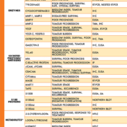
Melanoma cells release many proteins and other molecules into the extracellular fluid. Some of these molecules can end up in the bloodstream and hence serve as potential serum biomarkers. From a pathobiochemical point of view these biomarkers comprise molecules released by (i) necrotic cell content release, (ii) active secretion by melanoma cells, and (iii) ectodomain membrane shedding, including enzymes, soluble proteins/antigens, melanin-related metabolites, and circulating cell-free nucleic acids [1] [Table 1]. These molecules exhibit different prognostic and predictive values in melanoma diagnosis, staging, and treatment monitoring [1, 3–5].
Serum lactate dehydrogenase
In the American Joint Committee on Cancer (AJCC) staging system, serum lactate dehydrogenase (LDH) is the only serum biomarker that was accepted as a strong prognostic parameter in clinical routine for melanoma classifying those patients with elevated serum levels in stage IV M1C [3, 6].
Despite many promising results, there are also some limitations in measuring LDH as melanoma biomarker. First of all, LDH is not an actively secreted enzyme. Thus, LDH is only released through cell damage and cell death, which occur more frequently in malignant neoplasms. However, there are also false-positive values through hemolysis, hepatocellular injuries like hepatitis, myocardial infarction, muscle diseases, and other infectious diseases with high amounts of necrotic cells [3]. Moreover, LDH is non-specific for melanoma and elevated levels are also found in many other benign and malignant diseases.
Tyrosinase mRNA
An indicator for the presence of circulating melanoma cells and increased probability of the occurrence of metastases is the detection of tyrosinase mRNA in peripheral blood. Although the serological analyte is actually a nucleic acid isolated from circulating melanoma cells tyrosinase often is considered as an enzyme biomarker in melanoma [1, 3].
Due to the fact that tyrosinase mRNA is detected through nested RT-PCR the analytical sensitivity is very high. It is possible to detect one melanoma cell among 106 of normal blood cells. In recent decades, however, tyrosinase mRNA expression was determined in many different studies resulting in a wide range of variability (30–100%). One reason might be the transient presence of tumour cells in the bloodstream. On the other hand, non-standardized protocols for PCR-based techniques contribute to the observed variability, lower sensitivity, and different thresholds for melanoma cell detection.
Matrix metalloproteinases and cyclooxygenase-2
Further enzyme markers comprise matrix metalloproteinases and cyclooxygenase-2, with the latter detected via certain circulating eicosanoid products of the enzyme reaction [1, 7].
S100 calcium binding proteins
In addition, the S100 family of calcium binding proteins gained importance as both potential molecular key players and biomarkers in the etiology, progression, manifestation, and therapy of neoplastic disorders, including malignant melanoma. Moreover, S100 proteins receive attention as possible targets of therapeutic intervention moving closer to clinical impact.
In this regard, to-date, the best-studied S100 protein in melanoma is S100B [8, 9]. Increased S100B serum levels in melanoma patients chiefly have been attributed to the loss of cell integrity and proteolytic degradation as a result of apoptosis and necrosis of tumour cells. S100B seems to be the most promising serum marker for advanced melanoma, even more specific and sensitive than LDH, but is not yet applied in the clinical routine [1, 10].
Another member of the S100 family, the metastasis-associated protein S100A4 influences cell motility, angiogenesis, and apoptosis. The mechanism by which S100A4 stimulates metastasis is still under investigation; however, extracellular S100A4 seems to be of major importance in this context and, therefore, possibly might serve as a blood marker. Despite some early promising results on the use of S100A4 serum levels as a prognostic marker in melanoma, the greatest problem might be the low protein concentration in the blood which impedes clinical relevance [1]. This seems to be also true for other S100 proteins that are suggested to be biomarker candidates of melanoma. As more specific reagents for individual S100 proteins are being generated, their potential diagnostic and prognostic usage will increase substantially [1, 9].
Other candidate biomarkers
Other soluble proteins considered as melanoma biomarker candidates are given in Table 1. Furthermore, various non-protein biomarkers are potential targets for melanoma biomarker research. Those comprise metabolites of the melanin synthesis pathways, originating from the amino acid L-tyrosine, and cell-free nucleic acids [1].
Future directions in melanoma biomarker discovery
As well as the markers discussed above, other proteins, some of them possibly representing melanoma progenitor/stem cell-like markers, can be detected in circulating melanoma cells, at least as demonstrated in animal models. This includes ATP-binding cassette (ABC) multidrug transporters and neuroepithelial intermediate filament nestin [1, 5]. These markers offer the potential to predict the risk of progression to metastatic disease states, treatment resistance, and disease relapse. Lack of sufficient sensitivity, specificity, and accuracy are the most relevant limitations of a single blood-based melanoma biomarker for clinical use.
By contrast, a cluster of biomarkers for one disease would be a better diagnostic tool with much higher sensitivity, specificity, and clinical accuracy. Therefore, new investigations, called ´proteomic profiling´, focus on the identification of multiple co-expressed biomarkers or signature biomarker patterns, which allow early detection, staging, therapeutic monitoring and prognostic predictions [4, 11, 12].
Abbreviations
Biomarker abbreviations: 6H5MI2C, 6-hydroxy-5-methoxyindole-2-carboxylic acid; CEACAM, carcinoembryonic antigen-related cell adhesion molecule 1; CYT-MAA, cytoplasmic melanoma associated antigen; MAGE, melanoma associated antigen-1; MART-1, melanoma antigen recognized by T-cells 1; MIA, melanoma inhibitory activity; MMP, matrix metalloproteinase; sICAM, soluble intercellular adhesion molecule 1; sVCAM, soluble vascular cell adhesion molecule 1; TA90, tumour-associated antigen 90; VEGF, vascular endothelial growth factor; YKL-40, heparin- and chitin-binding lectin YKL-40 (syn. human cartilage glycoprotein-39)
Method abbreviations: ELISA, enzyme-linked immunosorbent assay; HPLC, high performance liquid chromatography; IHC, immunohistochemistry; IP, immunoprecipitation; LIA, luminescence immunoassay; RT-PCR, reverse transcription polymerase chain reaction; TMA, tissue microarray
This publication summarizes a comprehensive review article on protein and non-protein biomarkers in melanoma recently published by the authors [1, cf. references therein].
References
1. Tandler N, Mosch B, Pietzsch J. Protein and non-protein biomarkers in melanoma: a critical update. Amino Acids 2012; 43: 2203–2230.
2. De Giorgi V, Gori A, Grazzini M, et al. Epidemiology of melanoma: is it still epidemic? What is the role of the sun, sunbeds, Vit D, betablocks, and others? Dermatol Ther 2012; 25: 392–396.
3. Vereecken P, Cornelis F, Van Baren N, et al. A synopsis of serum biomarkers in cutaneous melanoma patients. Dermatol Res Pract 2012; 2012: 260643.
4. Palmer SR, Erickson LA, Ichetovkin I, et al. Circulating serologic and molecular biomarkers in malignant melanoma. Mayo Clin Proc 2011; 86: 981–990.
5. Mimeault M, Batra SK. Novel biomarkers and therapeutic targets for optimizing the therapeutic management of melanomas. World J Clin Oncol 2012; 3: 32–42.
6. Balch CM, Gershenwald JE, Soong SJ, et al. Final version of 2009 AJCC melanoma staging and classification. J Clin Oncol 2009; 27: 6199–6206.
7. Kruijff S, Hoekstra HJ. The current status of S-100B as a biomarker in melanoma. Eur J Surg Oncol 2012; 38: 281–285.
8. Nicolaou A, Estdale SE, Tsatmali M, et al. Prostaglandin production by melanocytic cells and the effect of alpha-melanocyte stimulating hormone. FEBS Lett 2004; 570: 223–226.
9. Pietzsch J. S100 proteins in health and disease. Amino Acids 2011; 41: 755–760.
10. Weide B, Elsässer M, Büttner P, et al. Serum markers lactate dehydrogenase and S100B predict independently disease outcome in melanoma patients with distant metastasis. Br J Cancer 2012; 107: 422–428.
11. Solassol J, Du-Thanh A, Maudelonde T, et al. Serum proteomic profiling reveals potential biomarkers for cutaneous malignant melanoma. Int J Biol Markers 2011; 26: 82–87.
12. Pham TV, Piersma SR, Oudgenoeg G, Jimenez CR. Label-free mass spectrometry-based proteomics for biomarker discovery and validation. Expert Rev Mol Diagn 2012; 12: 343–359.
Acknowledgements
Nadine Tandler is the recipient of a fellowship from the Europäische Sozialfonds (ESF).
The authors
Jens Pietzsch*, PhD, MD, Nadine Tandler, MSc and Birgit Mosch, PhD
Department of Radiopharmaceutical and Chemical Biology, Institute of Radiopharmaceutical Cancer Research, Helmholtz-Zentrum Dresden-Rossendorf, Dresden, Germany
*Corresponding author
E-mail: j.pietzsch@hzdr.de


