Researchers develop enzyme ‘atlas’ to help decipher cellular pathways
One of the most important classes of human enzymes are protein kinases – signalling molecules that regulate nearly all cellular activities, including growth, cell division, and metabolism. Dysfunction in these cellular pathways can lead to a variety of diseases, particularly cancer.
Identifying the protein kinases involved in cellular dysfunction and cancer development could yield many new drug targets, but for the vast majority of these kinases, scientists don’t have a clear picture of which cellular pathways they are involved in, or what their substrates are.
“We have a lot of sequencing data for cancer genomes, but what we’re missing is the largescale study of signalling pathway and protein kinase activation states in cancer. If we had that information, we would have a much better idea of how to drug particular tumors,” says Michael Yaffe, who is a David H. Koch Professor of Science at MIT, the director of the MIT Center for Precision Cancer Medicine, a member of MIT’s Koch Institute for Integrative Cancer Research, and one of the senior authors of the new study.
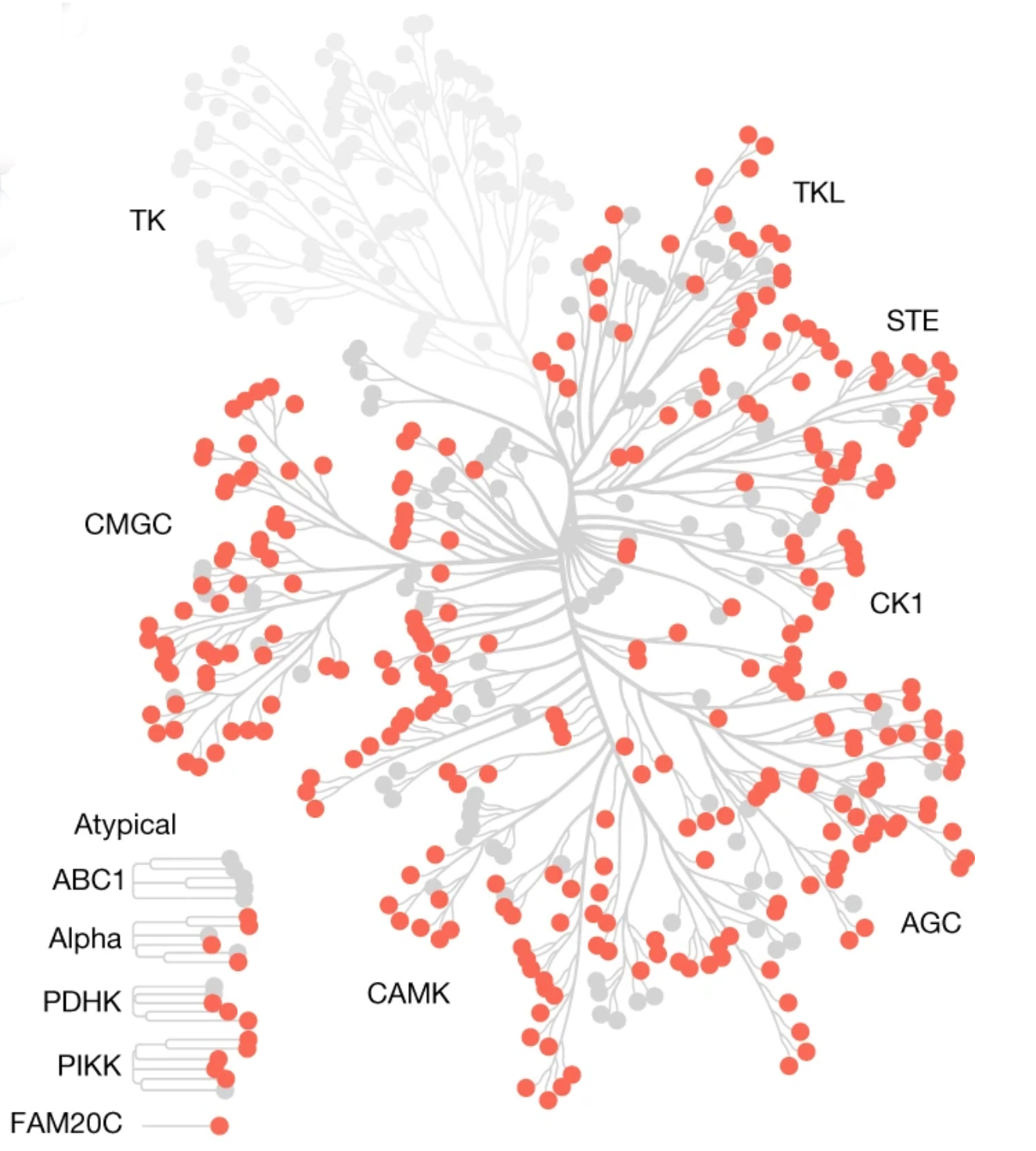
Yaffe and other researchers have now created a comprehensive atlas of more than 300 of the protein kinases found in human cells, and identified which proteins they likely target and control. This information could help scientists decipher many cellular signalling pathways, and help them to discover what happens to those pathways when cells become cancerous or are treated with specific drugs.
Lewis Cantley, a professor of cell biology at Harvard Medical School and Dana Farber Cancer Institute, and Benjamin Turk, an associate professor of pharmacology at Yale School of Medicine, are also senior authors of the paper, < https://doi.org/10.1038/s41586-022-05575-3 > which appears in Nature.
A Rosetta stone
The human genome includes more than 500 protein kinases, which activate or deactivate other proteins by tagging them with a chemical modification known as a phosphate group. For most of these kinases, the proteins they target are unknown, although research into kinases such as MEK and RAF, which are both involved in cellular pathways that control growth, has led to new cancer drugs that inhibit those kinases. To identify additional pathways that are dysregulated in cancer cells, researchers rely on phosphoproteomics using mass spectrometry to discover proteins that are more highly phosphorylated in cancer cells or healthy cells. However, until now, there has been no easy way to interrogate the mass spectrometry data to determine which protein kinases are responsible for phosphorylating those proteins. Because of that, it has remained unknown how those proteins are regulated or misregulated in disease.
“For most of the phosphopeptides that are measured, we don’t know where they fit in a signalling pathway. We don’t have a Rosetta stone that you could use to look at these peptides and say, this is the pathway that the data is telling us about,” Yaffe says. “The reason for this is that for most protein kinases, we don’t know what their substrates are.”
Twenty-five years ago, while a postdoc in Cantley’s lab, Yaffe began studying the role of protein kinases in signalling pathways. Turk joined the lab shortly after, and the three have since spent decades studying these enzymes in their own research groups.
“This is a collaboration that began when Ben and I were in Lew’s lab 25 years ago, and now it’s all finally really coming together, driven in large part by what the lead authors, Jared and Tomer, did,” Yaffe says.
In this study, the researchers analyzed two classes of kinases – serine kinases and threonine kinases, which make up about 85 percent of the protein kinases in the human body – based on what type of structural motif they put phosphate groups onto.
Working with a library of peptides that Cantley and Turk had previously created to search for motifs that kinases interact with, the researchers measured how the peptides interacted with all 303 of the known serine and threonine kinases. Using a computational model to analyze the interactions they observed, the researchers were able to identify the kinases capable of phosphorylating every one of the 90,000 known phosphorylation sites that have been reported in human cells, for those two classes of kinases.
To their surprise, the researchers found that many kinases with very different amino acid sequences have evolved to bind and phosphory-late the same motifs on their substrates. They also showed that about half of the kinases they studied target one of three major classes of motifs, while the remaining half are specific to one of about a dozen smaller classes.
Decoding networks
This new kinase atlas can help researchers identify signalling pathways that differ between normal and cancerous cells, or between treated and untreated cancer cells, Yaffe says.
“This atlas of kinase motifs now lets us decode signalling networks,” he says. “We can look at all those phosphorylated peptides, and we can map them back onto a specific kinase.”
To demonstrate this approach, the researchers analyzed cells treated with an anticancer drug that inhibits a kinase called Plk1, which regulates cell division. When they analyzed the expression of phosphorylated proteins, they found that many of those affected were controlled by Plk1, as they expected. To their surprise, they also discovered that this treatment increased the activity of two kinases that are involved in the cellular response to DNA damage.
Yaffe’s lab is now interested in using this atlas to try to find other dysfunctional signalling pathways that drive cancer development, particularly in certain types of cancer for which no genetic drivers have been found.
“We can now use phosphoproteomics to say, maybe in this patient’s tumour, these pathways are upregulated or these pathways are down-regulated,” he says. “It’s likely to identify signalling pathways that drive cancer in conditions where it isn’t obvious what the genetics that drives the cancer are.”


