Beukenlaan 137
5616 VD Eindhoven
The Netherlands
+31 85064 55 82
info@clinlabint.com
PanGlobal Media is not responsible for any error or omission that might occur in the electronic display of product or company data.
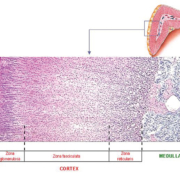
by Dr Petraki Munujos The detection of anti-adrenal cortex antibodies, also known as 21-hydroxylase or 21-OH antibodies, is an aid in the diagnosis and treatment of autoimmune adrenalitis. Far from being outdated, indirect immunofluorescence is a major analytical procedure used in the autoimmune laboratory for the measurement of these autoantibodies. Several techniques can be currently […]
Stroke biomarkers provide much insight into stroke biology that could be translated for patient benefit. When carefully harnessed, these biomarkers could guide decision-making in challenging clinical scenarios. This article offers an overview on current notable brain biomarkers that could aid clinicians in acute stroke management.
by Geelyn J.L. Ng and Dr Raymond C.S. Seet
Introduction
Stroke is a leading cause of permanent disability and the second most important cause of death globally [1]. Against a backdrop of a rapidly ageing society, there are concerns that a silent epidemic of stroke looms over our population.
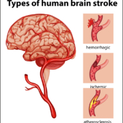
Ischemic stroke, a subset that affects 87% of stroke population, results from atherosclerosis that affects predominantly the cerebral vasculature. Atherosclerosis of the blood vessels can lead to a cessation or depletion of blood flow to the brain, triggering cerebral ischemia when brain tissues are no longer viable. Blood clots can also be formed in blood vessels and in the heart, subsequently dislodging into the brain (‘embolic stroke’). Presently, there are only two clinically adopted methods of acute reperfusion treatment – intravenous recombinant tissue plasminogen activator (TPA) [2] and endovascular treatment through device-driven retrieval or aspiration of blood clots [3]. Although good functional recovery is five times more likely to occur with early reperfusion [3], the use of acute reperfusion treatment is restricted to a small group of patients where the benefits of treatment are weighed against the persisting risk of hemorrhagic transformation [4].
Sieving out stroke patients who are at risk of recurrent attacks is the first step to enable accurate triaging of patients to specialized units for in-depth observation and individualized treatment for complications arising from stroke. Presently, such identification is highly reliant on a clinician’s intuition and knowledge of neurologic deficits, as well as neuroimaging results. Tapping into the use of cerebral ischemia biomarkers could shed light on the complex pathological consequences following ischemic stroke and bring forth an unbiased system to weigh risks and benefits of treatments for clinicians and researchers alike.
Biomarkers are biological indicators of physiology that are objectively measured for use in risk stratification and development of therapeutic strategies. Having high sensitivity and specificity for the outcome it is expected to diagnose is generally a trait of a good biomarker, especially when targeting a complex and heterogenous disease such as stroke. Using a multi-biomarker platform could aim at different pathways of this multifaceted disease, thereby allowing for a more comprehensive treatment. Due to the presence of the blood-brain barrier (BBB) that holds a tight control over the inflow and outflow of particles, human brain tissues are typically difficult to access, making it impracticable to measure a biomarker within the brain. During cerebral ischemia, the BBB is broken down, causing brain-derived biomarkers to be released into the blood circulation, making it possible for a closer examination of the pathologic processes that take place following stroke onset. Although many biomarkers exist that could aid in stroke research, we have previously focused on notable blood-based stroke biomarkers that may play a bigger part in supporting the difficult clinical decision-making process [5]. This article will be providing an overview of several well-researched blood-based biomarkers, with much potential in aiding the clinical assessment of stroke patients.
Stroke biomarkers in the clinical scene
Studies in ischemic stroke have investigated the usefulness of blood-based biomarkers in identifying stroke mimics, establishing stroke etiology and prognosticating stroke severity and outcomes, such as vascular events and functional recovery [6, 7]. Presently, use of biomarkers in routine clinical practice remains uncommon, as stroke severity is still determined mainly through a thorough clinical neurological assessment and subjective interpretation of neuroimaging findings by a skilled physician. Nevertheless, having an objective means to prognosticate an outcome via a blood sample retrieved from a patient upon stroke presentation could add value to clinical decision-making, especially during times when neuroimaging results and clinical interpretations are unable to yield conclusive results. As stroke is a heterogeneous condition, investigating biomarkers that target different stroke pathways could be promising in establishing a multi-biomarker platform, especially for outcomes such as hemorrhagic transformation (HT), early neurologic deterioration (END) and malignant cerebral infarction.
Matrix metalloproteinase-9
Although administrating TPA could potentially achieve the benefit of arterial recanalization, the risk of symptomatic intracranial secondary hemorrhage within the infarcted brain tissues must not be forgotten. Matrix metalloproteinase-9 (MMP-9) is an enzyme that degrades the basal lamina and breaks down the extracellular matrix when activated during TPA treatment. The function of the BBB is crippled in this process and an inflammatory cascade is initiated, resulting in edema and the dreaded HT [8]. Apart from its involvement in HT, MMP-9 could also be used to identify high-risk END patients and plays a part in malignant cerebral infarction.
C-reactive protein
C-reactive protein (CRP) is a sensitive systemic marker of inflammation and a well-researched biomarker of ischemic stroke found in the blood plasma. CRP has been associated with END and noted to be predictive of adverse outcome, where ischemic stroke patients with higher CRP levels tend to suffer from a significantly worse outcome and mortality [9, 10].
S100β
S100β is a biomarker of ischemic stroke expressed by neuronal cells that can be released into the bloodstream when the BBB is compromised. Its concentration needs to be carefully balanced, as it may be protective in low concentrations, but at high levels has been shown to predict cerebral malignant infarction and correlate with infarct size [11, 12]. However, trial data on the use of biomarkers to guide clinical decisions leading to early decompressive surgery are currently lacking. Several studies have also uncovered an increase in S100β in ischemic stroke patients who present 1 to 7 days from symptom onset [13, 14]. In acute stroke patients, elevated S100β serum levels before thrombolytic therapy have also been demonstrated as a risk factor for HT [15].
N-terminal pro-brain natriuretic peptide
The brain natriuretic peptide (BNP) and its precursor, N-terminal proBNP (NT-proBNP), have been extensively studied as useful biomarkers for the prognosis and diagnosis of heart failure [16]. In recent years, BNP is gradually gaining recognition as a marker of atrial fibrillation (AF) and, therefore, as a biomarker to diagnose and predict stroke of cardioembolic origin [17, 18]. Plasma BNP levels have also been demonstrated to have significant correlations with infarct volume and National Institutes of Health stroke scale (NIHSS), making it a potentially powerful clinical biomarker for acute ischemic stroke [19].
Uric acid
Although uric acid has been adopted clinically for metabolic diseases, it is slowly garnering interest in the field of cardiovascular diseases due to its antioxidant properties. Despite data to suggest a strong association between uric acid levels and positive stroke outcomes [20, 21], several studies have observed an adverse relationship where higher uric acid levels were found to predict poor functional outcome and increased mortality [22, 23]. This disparity could highlight a dual role of uric acid in stroke, where both high and low levels of uric acid could adversely affect stroke outcomes [24]. Much remains to be explored for this biomarker before it could be roled out for use in stroke prognosis or diagnosis.
F2-isoprostanes
The product of arachidonic acid peroxidation generated by free radicals, F2-isoprostanes is well-established as a reliable biomarker for oxidative damage. Even though stroke is widely known as partly the result of oxidative damage, the relationship between F2-isoprostanes and human stroke remains poorly understood. Studies have demonstrated its importance in ischemic stroke as elevated levels of F2-isoprostanes could be observed in patients during the early course of stroke onset, with one even as early as three hours after [25–27].
Conclusion
Cerebral ischemia biomarkers have the potential to bridge translational gaps in medicine by shedding light on the pathological events leading to cerebral infarction and the ischemic cascade, aiding in clinical assessment during the critical time-sensitive decision-making process. Results in this area are still emerging, and more efforts could focus on ensuring the feasibility of incorporating stroke biomarkers for patient benefit. The translation of stroke biomarkers to clinical practice is challenging but can be extremely rewarding, especially when such concerted efforts of researchers, clinicians, industry partners and regulatory authorities result in a positive outcome for stroke patients.
Acknowledgements
We would like to thank the National Medical Research Council, Singapore (NMRC/CSA-SI/0003/2015, NMRC/CNIG/1115/2014 and NMRC/MOHIAFCat1/0015/2014) for their generous support.
References
1. GBD 2015 Mortality and Causes of Death Collaborators. Global, regional, and national life expectancy, all-cause mortality, and cause-specific mortality for 249 causes of death, 1980-2015: a systematic analysis for the Global Burden of Disease Study 2015. Lancet 2016; 388(10053): 1459–1544.
2. NINDS rt-PA Stroke Study Group. Tissue plasminogen activator for acute ischemic stroke. N Engl J Med 1995; 333(24): 1581–1587.
3. Powers WJ, Derdeyn CP, Biller J, Coffey CS, Hoh BL, Jauch EC, et al. 2015 American Heart Association/American Stroke Association focused update of the 2013 guidelines for the early management of patients with acute ischemic stroke regarding endovascular treatment: a guideline for healthcare professionals from the American Heart Association/American Stroke Association. Stroke 2015; 46(10): 3020–3035.
4. Seet RC, Rabinstein AA. Symptomatic intracranial hemorrhage following intravenous thrombolysis for acute ischemic stroke: a critical review of case definitions. Cerebrovasc Dis 2012; 34(2): 106–114.
5. Ng GJL, Quek AML, Cheung C, Arumugam TV, Seet RCS. Stroke biomarkers in clinical practice: a critical appraisal. Neurochem Int 2017; 107: 11–22.
6. Bustamante A, López-Cancio E, Pich S, Penalba A, Giralt D, García-Berrocoso T, et al. Blood biomarkers for the early diagnosis of stroke: The Stroke-Chip Study. Stroke 2017; 48(9): 2419–2425.
7. Whiteley W, Chong WL, Sengupta A, Sandercock P. Blood markers for the prognosis of ischemic stroke: a systematic review. Stroke 2009; 40(5): e380–389.
8. Barr TL, Latour LL, Lee KY, Schaewe TJ, Luby M, Chang GS, et al. Blood-brain barrier disruption in humans is independently associated with increased matrix metalloproteinase-9. Stroke 2010; 41(3): e123–128.
9. Muir KW, Weir CJ, Alwan W, Squire IB, Lees KR. C-reactive protein and outcome after ischemic stroke. Stroke 1999; 30(5): 981–985.
10. Idicula TT, Brogger J, Naess H, Waje-Andreassen U, Thomassen L. Admission C-reactive protein after acute ischemic stroke is associated with stroke severity and mortality: the ‘Bergen stroke study’. BMC Neurol 2009; 9: 18.
11. Vahedi K, Hofmeijer J, Juettler E, Vicaut E, George B, Algra A, et al. Early decompressive surgery in malignant infarction of the middle cerebral artery: a pooled analysis of three randomised controlled trials. Lancet Neurol 2007; 6(3): 215–222.
12. Abraha HD, Butterworth RJ, Bath PM, Wassif WS, Garthwaite J, Sherwood RA. Serum S-100 protein, relationship to clinical outcome in acute stroke. Ann Clin Biochem 1997; 34(Pt4): 366–370.
13. Aurell A, Rosengren LE, Karlsson B, Olsson JE, Zbornikova V, Haglid KG. Determination of S-100 and glial fibrillary acidic protein concentrations in cerebrospinal fluid after brain infarction. Stroke 1991; 22(10): 1254–1258.
14. Buttner T, Weyers S, Postert T, Sprengelmeyer R, Kuhn W. S-100 protein: serum marker of focal brain damage after ischemic territorial MCA infarction. Stroke 1997; 28(10): 1961–1965.
15. Foerch C, Wunderlich MT, Dvorak F, Humpich M, Kahles T, Goertler M, et al. Elevated serum S100B levels indicate a higher risk of hemorrhagic transformation after thrombolytic therapy in acute stroke. Stroke 2007; 38(9): 2491–2495.
16. Yancy CW, Jessup M, Bozkurt B, Butler J, Casey DE, Drazner MH, et al. 2013 ACCF/AHA Guideline for the management of heart failure: A report of the American College of Cardiology Foundation/American Heart Association Task Force on practice guidelines. Circulation 2013; 128(16): e240–327.
17. Naya T, Yukiiri K, Hosomi N, Takahashi T, Ohkita H, Mukai M, et al. Brain natriuretic peptide as a surrogate marker for cardioembolic stroke with paroxysmal atrial fibrillation. Cerebrovasc Dis 2008; 26(4): 434–440.
18. Fonseca AC, Matias JS, Pinho e Melo T, Falcao F, Canhao P, Ferro JM. N-terminal probrain natriuretic peptide as a biomarker of cardioembolic stroke. Int J Stroke 2011; 6(5): 398–403.
19. Tomita H, Metoki N, Saitoh G, Ashitate T, Echizen T, Katoh C, et al. Elevated plasma brain natriuretic peptide levels independent of heart disease in acute ischemic stroke: correlation with stroke severity. Hypertens Res 2008; 31(9): 1695–1702.
20. Amaro S, Urra X, Gomez-Choco M, Obach V, Cervera A, Vargas M, et al. Uric acid levels are relevant in patients with stroke treated with thrombolysis. Stroke 2011; 42(1 Suppl): S28–32.
21. Chamorro A, Obach V, Cervera A, Revilla M, Deulofeu R, Aponte JH. Prognostic significance of uric acid serum concentration in patients with acute ischemic stroke. Stroke 2002; 33(4): 1048–1052.
22. Weir CJ, Muir SW, Walters MR, Lees KR. Serum urate as an independent predictor of poor outcome and future vascular events after acute stroke. Stroke 2003; 34(8): 1951–1956.
23. Karagiannis A, Mikhailidis DP, Tziomalos K, Sileli M, Savvatianos S, Kakafika A, et al. Serum uric acid as an independent predictor of early death after acute stroke. Circ J 2007; 71(7): 1120–1127.
24. Seet RC, Kasiman K, Gruber J, Tang SY, Wong MC, Chang HM, et al. Is uric acid protective or deleterious in acute ischemic stroke? A prospective cohort study. Atherosclerosis 2010; 209(1): 215–219.
25. Sanchez-Moreno C, Dashe JF, Scott T, Thaler D, Folstein MF, Martin A. Decreased levels of plasma vitamin C and increased concentrations of inflammatory and oxidative stress markers after stroke. Stroke 2004; 35(1): 163–168.
26. Ward NC, Croft KD, Blacker D, Hankey GJ, Barden A, Mori TA, et al. Cytochrome P450 metabolites of arachidonic acid are elevated in stroke patients compared with healthy controls. Clin Sci (Lond) 2011; 121(11): 501–507.
27. Seet RC, Lee CY, Chan BP, Sharma VK, Teoh HL, Venketasubramanian N, et al. Oxidative damage in ischemic stroke revealed using multiple biomarkers. Stroke 2011; 42(8): 2326–2329.
The authors
Geelyn J.L. Ng1,2 BSc, Raymond C.S. Seet*1,2 MBBS, MRCP (UK), MMed (Int Med), FRCP (UK)
1Department of Medicine, Yong Loo Lin School of Medicine, National University of Singapore, Singapore
2Division of Neurology, Department of Medicine, National
University Health System, Singapore
*Corresponding author
E-mail: raymond_seet@nuhs.edu.sg
The need to differentiate patients with advanced liver disease from those with earlier stage, or more benign diseases for optimal management and allocation of resources is an ever present challenge. In this article we discuss our experiences of using the aspartate transaminase (AST) : alanine transaminase (ALT) ratio as part of a pathway to screen patients for referral to secondary care.
by Dr Raphael Buttigieg and Dr Sara Jenks
Introduction
Deaths from liver disease in Scotland are on the increase [1]. More often than not patients are picked up at a late stage of their disease with significant fibrosis and/or cirrhosis already present. As a result there is a need to try to identify patients with progressive disease earlier on in the course of their illness.
Abnormal liver function tests (LFTs) are frequently picked up on general screening blood samples done in primary care. The degree of abnormality correlates poorly with the extent of liver disease. The gold standard test for liver disease diagnosis and staging is considered to be a liver biopsy; however, there are many other considerations to this invasive procedure including clinical risk, technical ability of person doing the biopsy, inter-pathologist variation in scoring and others. These limitations have led to the development of non-invasive methods for the assessment of liver fibrosis. Although there have been suggestions by different groups regarding the appropriate use of non-invasive fibrosis scoring systems, no one guideline is currently in use.
Non-invasive methods rely on two different approaches [2]:
(a) A biomarker-based approach using serum samples. Advantages are their high applicability (>95%) and good inter-laboratory reproducibility.
(b) A physical approach based on the measurement of liver stiffness. Liver stiffness corresponds to an intrinsic physical property of liver parenchyma. Physical approaches include transient elastography such as FibroScan® and MR elastography.
Because of noted variations in care, as well as to ensure appropriate referrals, NHS Lothian made a guideline for GPs in 2013. At the time, based on the best available clinical evidence, the aspartate transaminase (AST) : alanine transaminase (ALT) ratio was chosen as a scoring system to guide referrals, which was developed in recognition that as liver fibrosis develops, the normal ratio tends to reverse. An abnormal AST:ALT ratio can, thus, be used to pick up patients who should be referred to secondary care for further investigation, as well as closer monitoring and treatment.
However, other biomarker-based fibrosis risk scores have also been developed [2], which have been used for this purpose including the Fibrosis-4 (FIB-4) [3], NAFLD (non-alcoholic fatty liver disease) fibrosis score, and APRI (AST to platelet ratio index), which may have a better performance than the AST:ALT ratio [4]. Each of these has been validated for different liver diseases – and in many cases different cut-off points are recommended for diagnosis of advancing fibrosis based on the likely primary pathology involved in the individual patient. For example, alcohol use in itself will raise the AST and, thus, the same AST:ALT ratio is likely to indicate more advanced fibrosis in someone with HCV-related liver disease than in alcoholic liver disease with ongoing ethanol excess. This adds to the complexity of using any one score in a guideline to ensure the right balance between sensitivity and specificity.
A final consideration to note is that specifically for NAFLD/non-alcoholic steatohepatitis (NASH), the continued development of pharmaceuticals for the prevention of disease progression means that, once again, the threshold for diagnosis may need to change as therapies to target earlier stages become available [5].
This article will discuss our guideline (Fig. 1) and conclusions drawn from an audit of its use.
Method
A list of all the requests for a AST:ALT ratio in a 6-month period in NHS Lothian was obtained from laboratory records – in terms of date of request, patient name and CHI number (unique patient identifier) (n=874). These records were encoded into a spreadsheet and a plan for analysis made.
Following this, various data were audited retrospectively from the patient electronic record. Individual notes and files were not used because of the logistical difficulty in analysing large numbers of case notes.
Of the total number of ratios (n=874) requested in the 6-month period, 49 were elevated at >1.0 and 295 were normal at ≤1.0; 530 ratio requests were cancelled due to ALT being within the reference range.
Results
The various aspects of the referral process from primary to secondary care were audited with the following aims.
1. To identify all the abnormal ratios in a 6-month period (n=49) (Table 1).
2. To identify all the ratios in the same 6-month period that were in the range 0.8–1.0 (n=53) (Table 2).
This was carried out to assess whether there should be concern about the ratio producing false negative results, and how useful it was to actually exclude liver disease. We thus audited all patients with a borderline ratio of 0.8–1.0.
Additionally, we asked if the FIB-4 score or APRI score was used, would this have affected referral?
3. To identify the first 50 individuals in a 6-month period tested with an ALT level of 40–49 on whom the AST:ALT ratio had been cancelled (n=50) (Table 3).
Although an upper limit of 50 is taken for the normal range of ALT, there is evidence that even at levels below this a certain amount of liver inflammation is present, and, thus, different health boards use other values – such as an upper limit of normal of 40.
This last part of the project set out to identify people with a borderline abnormal ALT of 40–49, and assess whether using different scores – such as the FIB-4 or APRI scores would potentially label these individuals as having liver disease and needing to be referred
Limitations of our study
Most of these patients had a very short-term follow-up, which in many cases did not allow proper determination of their disease severity, as well as assessment of long-term mortality/morbidity risk using different scoring systems.
Secondly, we were unable to compare scores to a gold standard as in many cases a liver biopsy had not been carried out. Transient elastography and hyaluronic acid testing had been carried out in a selection of patients which allowed further characterization of fibrosis staging; however, it is appreciated that neither of these are the gold standard.
Conclusions and considerations
The AST:ALT ratio is a good test for assessing whether people should be referred to secondary care or not. This conclusion is based on the fact that many patients who were referred with a positive ratio were seen in secondary care and kept under review. However, better tests are needed to further assess their stage of disease, ideally non-invasively.
The FIB-4 (possibly in association with further tests below) may be a more sensitive/specific score to be used in diagnosing patients; however, cut-off points would need to be determined to guide the most effective use of available resources in primary and secondary care. As can be seen in the second group FIB-4 and the APRI were raised in patients which would not have been picked up by the AST:ALT ratio, which thus increases pick-up. Another consideration is, as previously mentioned, that the AST:ALT ratio tends to be raised in patients drinking excessive ethanol, even if their disease is not very advanced. Since in our cohort alcohol use was very prevalent, other scores may possibly be better suited.
The plan from now is to adopt a pathway of cascading lab tests based on patients’ alcohol consumption, BMI/metabolic syndrome markers, LFT results and automatic scoring with interpretation will be issued to GPs. Also possible is further testing – either in the community or in secondary care to further guide patients in different scoring groups – including either transient elastography (FibroScan), or further biochemical testing. NHS Lothian currently uses hyaluronic acid, and this may be a way of further classifying/evaluating people in ‘intermediate’ categories. The elastography (FibroScan) test could be another option and this is the current recommendation in the current NICE guidelines.
For any further information please feel free to contact the authors:
Raphael Buttigieg: ST3 Chemical Pathology and Metabolic Medicine, NHS Greater Glasgow and Clyde, UK; raphael.buttigieg@nhs.net.
Sara Jenks: Consultant in Chemical Pathology and Metabolic Medicine, NHS Lothian, Edinburgh, UK; sjenks@nhs.net.
References
1. Gray L, Leyland AH. Alcohol. The Scottish Health Survey 2014: Volume 1: Main report (http://www.gov.scot/Publications/2015/09/6648/318753)
2. European Association for Study of Liver. EASL-ALEH Clinical Practice Guidelines: Non-invasive tests for evaluation of liver disease severity and prognosis. J Hepatol 2015; 63(1): 237.
3. McPherson S, Anstee QM, Henderson E, Day CP, Burt AD. Are simple noninvasive scoring systems for fibrosis reliable in patients with NAFLD and normal ALT levels? Eur J Gastroenterol Hepatol 2013; 25(6): 652–658.
4. Parkes J, Guha IN, Harris S, Rosenberg WM, Roderick PJ. Systematic review of the diagnostic performance of serum markers of liver fibrosis in alcoholic liver disease. Comp Hepatol 2012; 11(1): 5.
5. Dyson JK, Anstee QM, McPherson S. Non-alcoholic fatty liver disease: a practical approach to diagnosis and staging. Frontline gastroenterology 2014; 5(3):211–218.
The authors
Raphael Buttigieg*1 Sara Jenks2
1Department of Clinical biochemistry, Glasgow Royal Infirmary, NHS Greater Glasgow and Clyde, UK
2Department of Clinical biochemistry, NHS Lothian, Royal Infirmary of Edinburgh, Edinburgh, UK
*Corresponding author
E-mail: raphael.buttigieg@nhs.net
by Dr Jacqueline Gosink The international consensus on standardized nomenclature of human epithelial cell (HEp-2 cell) patterns in indirect immunofluorescence (ICAP, www.anapatterns.org) defines fifteen nuclear patterns, nine cytoplasmic patterns and five mitotic patterns which are relevant for the diagnosis of various autoimmune diseases. Furthermore, the consensus stipulates that autoantibodies detected by indirect immunofluorescence on HEp-2 […]
Mild traumatic brain injury (mTBI) and concussion from sporting/recreational activities are relatively common. However, assessment of mTBI is difficult and many incidents of mTBI and concussion are unrecognized and/or not reported. Levels of the α-amino-3-hydroxy-5-methyl-4-isoxazolepropionic acid receptor (AMPAR) peptide, a product of the proteolytic degradation of AMPA receptors, have been found to be raised in mTBI and the development of point-of-care (POC) tests based on the recognition of the AMPAR peptide is underway. Such POC tests will be useful at the pitch sideline or in the combat field for aiding objective diagnosis and management of subtle brain injury.
by Prof. Svetlana A. Dambinova, Rozalyn Heath and Dr Galina A. Izykenova
Introduction
Mild traumatic brain injury (mTBI) including concussion is the most frequent form of injury in military and civilian settings with the highest prevalence among young adults aged 15 to 24 years. Each year sports and recreational activities contribute about 3.8 million cases of mTBI in the USA [1], whereas brain injuries caused by explosions are the most common combat wounds in the military arena.
Assessment of mTBI regardless of origin is complicated. Many primary mTBIs, and particularly concussions, go unrecognized or are not reported when there is no loss of consciousness. Additionally, without sufficient reports of previous incidents, soldiers and competitive athletes are often subjected to multiple concussions. There are several challenges to identify immediate (or primary acute and subacute within 24 hours and up to 2 weeks respectively) impact, secondary (beyond 14 days) and cumulative (brain-related seizures) consequences that might follow multiple concussions or mTBI.
Normally, acute subclinical concussions associated with micro-edema formation that is reversible and not visible on conventional computed tomography and magnetic resonance imaging (MRI) methods. Advanced neuroimaging techniques (diffusion tensor imaging, functional MRI, and positron emission tomography) that can register minor structural and microvascular changes are primarily used for research. These modalities are not available in emergency situations or for routine clinical evaluations and have a limited application in persons with metal implants [2] or claustrophobia [3].
Currently, there is an unmet diagnostic need to reveal brain micro-damage following concussion by use of a rapid and affordable assay detecting, for instance, neurotoxicity (immunoexcitotoxicity) biomarkers in the bloodstream [4]. Analogous to NR2 peptide as a biomarker for cortical lesions in transient ischemic attack (TIA)/strokes [5], we proposed that the α-amino-3-hydroxy-5-methyl-4-isoxazolepropionic acid receptor (AMPAR) peptide marker is able to differentiate subtle brain injury in white matter associated with concussions.
Neurotoxicity biomarkers in mTBI
It is known that the family of ionotropic glutamate receptors (GluRs) implying N-methyl-D-aspartate (NMDAR), AMPAR and kainite receptors are involved in the regulation of synaptic connectivity in cortical/subcortical and brainstem areas [4,5]. Recently, it was shown that AMPAR represents a biomarker for the neurotoxicity cascade underlying subtle brain injury [6].
The family of location-specific GluRs is involved in more than 80% of cortical and subcortical neuronal communications underlying superior mental functions [7]. AMPAR is primarily distributed in the forebrain and subcortical pathways [8], and strategically located on surfaces of small cerebral arteries regulating blood circulations in white matter substructures [9]. Symptomatically the impact to brain may lead to executive brain dysfunctions associated with subcortical areas. Visual and cognitive deficits (including problems with memory, intellect, concentration and attention) might be an aftermath of subtle repetitive injuries to deep brain structures due to more severe cases of mTBI.
In acute and subacute phases of mTBI, a massive release of glutamate, which upregulates excitotoxic AMPARs has been detected [4]. The GluR1-subunit of N-terminal AMPAR fragments is rapidly cleaved by extracellular proteases and fragments carrying immune active epitopes released into the bloodstream through the compromised blood–brain barrier (BBB). This degradation product can be detected directly in the blood as AMPAR peptide fragments (molecular weight 5–7 kDa) [4]. The protective effects of a compromised BBB impacting neurotoxicity are exacerbated further when accompanied by a delayed immunological response generating peripheral anti-CNS antibodies [10].
Concussion assays development
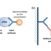
To detect AMPAR peptide, magnetic-particle-based enzyme-linked immunosorbent assay (MP-ELISA) containing unique reagents has been developed. MP-ELISA involved a sandwich or ‘bridging’ assay where the suspended in solution microparticles coated by capture antibodies are binding to two epitopes of AMPAR peptide and reaction is revealed by probe-detection antibodies (Fig. 1). The probe is an enzyme that generates a colour reaction. The assay includes control samples (reference standards) produced synthetically or as a fusion human protein.
A feasibility study detecting the AMPAR peptide in a single blood draw taken from club sport athletes and professional football players in acute and subacute stage of concussions evaluated cut-offs of 0.4 and 1.0 ng/mL respectively for the assessment of single and recurrent concussions (Table 1) [11,12]. The predictive value of the test was assessed as 91% with a likelihood ratio of 11–12 for recognizing individuals with mTBI. If the test at a cut-off point of 0.4 ng/mL was negative, the post-test probability for a single concussion would be <4%.
AMPAR peptide concentrations in plasma for a military cohort suffering mTBI showed increased levels with an average concentration of 2.98 ng/mL [13]. In this study, the optimal cut-off value for recurrent mTBI was similar to professional players (Table 1), at which a positive predictive value of 93% was achieved. The trade-offs between true-positive and false-positive yielded in an area under the receiver operating characteristic (ROC) curve of 0.97.
Early experimental and clinical research of antibodies to AMPA receptors (AMPAR Ab) as an immunoexcitotoxicity biomarker has demonstrated their diagnostic value in detecting pathological brain-spiking activity and epileptic seizures [14,15] as a consequence of traumatic brain injury, thereby representing a prognostic risk factor [16]. Clinical studies of GluR1 antibodies in adult patients with different chronic neurological pathology (n=1866) performed in Russia, Germany, Ireland, Poland and the USA have demonstrated diagnostic potential (sensitivity of 86%-88% and specificity of 83–97%) in assessment of post-traumatic seizures.
In sport-related multiple concussions, AMPAR Ab values remained abnormally high in the blood of some athletes who had headaches and visual problems. It was suggested that this finding may reflect persistent changes in the subcortical areas of the brain. The diagnostic value of AMPAR Ab (sensitivity of 86–88%, specificity of 83–97% at 1.5 ng/ml cut-off) in assessment of seizures defined by electroencephalogram (EEG) with history of sustained single or multiple TBI have been demonstrated for children and adult patients indicating a development of ‘chronic’ conditions [13].
Sideline testing of AMPAR peptide and antibody
Recognizing the medical need for sideline testing for mTBI (largely for emergency care), a point-of-care (POC) testing platform has been recently undertaken for AMPAR peptide and antibody assays are being tested in clinical studies (www.drdbiotech.com).
A lateral flow sandwich assay to detect AMPAR Ab consists of a blood filtering sample pad, a pad containing gold nanoshells conjugated to protein A, a nitrocellulose strip with immobilized AMPAR peptide, a control line, and a cellulose absorbent. Applied to the blood filter, erythrocytes are removed from the sample, which passes to the conjugate pad where AMPAR IgGs are captured by protein A. When the complex reaches the peptide test line stripe on nitrocellulose, it binds to the test line yielding a visible signal with intensity proportional to the concentration of the AMPAR Ab presented in the sample (Fig. 2).
The control line then captures a portion of the remaining nanoshells regardless of the presence or absence of the peptide (Fig. 3).
The AMPAR peptide prototype test that works on a similar principle, employed gold nanoparticles as the signal-generating species covalently bound to specific antibodies against AMPAR peptide. This assay captures the AMPAR peptide from the blood sample between two different Abs, one immobilized on the nitrocellulose and the other on the gold nanoshells. This ‘bridging’ of the analyte leads to the immobilization of the particles at the test line producing a colour signal.
Conclusion
The laboratory assessment of concussion at the pitch sideline in competitive contact sports or on the field of combat is required to assist in the objective confirmation of when an injured athlete should return to play or a soldier may return to duty. Accurate diagnosis of concussion and appropriate management of subtle brain injury to prevent damage to the long-term health of athletes and others at risk of re-injury. Specific brain biomarkers detected by a rapid blood test would have important diagnostic/prognostic capabilities, particularly for concussion, to objectively evaluate early signs of further brain-function deterioration and assist in navigating personalized therapy when required. Clinical use of blood tests can reliably detect subtle brain impact, predict consequences and aid in triaging persons with persistent symptoms after a recurrent concussion for diffuse tensor or diffuse-weighted imaging modalities of MRI [17].
Early identification of concussion has the potential to become a key component of a successful treatment strategy and outcome monitoring. Advances in analytical assay technologies have made it possible to develop a rapid, cost-effective test that can be used to target populations and select a risk group for post-traumatic seizures to direct to the immediate attention of a specialist.
The opportunity of using ‘yes/no’ lateral flow tests without a need for an instrumental readout, being simply read by eye, would make a huge difference in supplying coaches with reliable, simple and rapid tests in return-to-play decisions. There is also a high demand for portable POC tests for mTBI diagnosis in military combat and civilian emergency settings.
References
1. Faul M, Xu L, Wald MM, Coronado VG. Traumatic brain injury in the United States. Emergency department visits, hospitalizations, and deaths 2002–2006. Atlanta, GA. Centers for Disease Control and Prevention, National Center for Injury Prevention and Control 2010 (https://www.cdc.gov/traumaticbraininjury/pdf/blue_book.pdf).
2. Klinke T, Daboul A, Maron J, Gredes T, Puls R, Jaghsi A, Biffar R. Artifacts in magnetic resonance imaging and computed tomography caused by dental materials. PLoS One 2012; 7: e31766–31771.
3. Napp AE, Enders J, Roehle R, Diederichs G, Rief M, Zimmermann E, Martus P, Dewey M. Analysis and prediction of claustrophobia during MR imaging with the Claustrophobia Questionnaire: an observational prospective 18-month single-center study of 6500 patients. Radiology 2017; 283: 148–157.
4. Dambinova SA. Diagnostic challenges in traumatic brain injury. IVD Technology 2007; 3: 3–7.
5. Dambinova SA. Biomarkers for transient ischemic attack (TIA) and ischemic stroke. Clin Lab Inter 2008; 32: 7–10.
6. Danilenko UD, Khunteev GA, Bagumyan A, Izykenova GA. Neurotoxicity biomarkers in experimental acute and chronic brain injury. In: Dambinova SA, Hayes RL, Wang KKW (editors). Biomarkers for TBI. RSC Publishing, RSC Drug Discovery Series 2012; pp87–98.
7. Gill S, Pulido O. Glutamate receptors in peripheral tissue. In: Gill S, Pulido O (editors). Excitatory transmission outside the CNS. Kluwer Academic Publishers 2010; p3.
8. Hammond JC, McCullumsmith RE, Funk AJ, Haroutunian V, Meador-Woodruff JH. Evidence for abnormal forward trafficking of AMPA receptors in frontal cortex of elderly patients with schizophrenia. Neuropsychopharmacology 2010; 35: 2110–2119.
9. Christensen PC, Samadi-Bahrami Z, Pavlov V, Stys PK, Moore GR. Ionotropic glutamate receptor expression in human white matter. Neurosci Lett 2016; 630: 1–8.
10. Raad M, Nohra E, Chams N, Itani M, Talih F, Mondello S, Kobeissy F. Autoantibodies in traumatic brain injury and central nervous system trauma. Neuroscience 2014; 281: 16–23.
11. Dambinova SA, Shikuev AV, Weissman JD, Mullins JD. AMPAR peptide values in blood of nonathletes and club sport athletes with concussions. Mil Med 2013; 3: 285–290.
12. Dambinova SA, Maroon JC, Sufrinko AM, Mullins JD, Alexandrova EV, Potapov AA. Functional, structural, and neurotoxicity biomarkers in integrative assessment of concussions. Front Neurol 2016; 7: 172.
13. Mullins JD. Biomarkers of TBI: implications for diagnosis and management of contusions. AMSUS 118th Annual Continuing Education Meeting. Seattle, WA, USA 2013; 147. (http://amsusce.org/wp-content/uploads/2015/05/Abstract-Summaries-10.22.13.2.pdf)
14. Dambinova SA, Izykenova GA, Burov SV, Grigorenko EV, Gromov SA. The presence of autoantibodies to N-terminus domain of GluR1 subunit of AMPA receptor in the blood serum of patients with epilepsy. J Neurol Sci 1997; 152: 93–97.
15. Dambinova SA, Granstrem OK, Tourov A, Salluzzo R, Castello F, Izykenova GA. Monitoring of brain spiking activity and autoantibodies to N-terminus domain of GluR1 subunit of AMPA receptors in blood serum of rats with cobalt-induced epilepsy. J Neurochem 1998; 71: 2088–2093.
16. Goryunova AV, Bazarnaya NA, Sorokina EG, Semenova NY, Globa OV, Semenova ZhB, Pinelis VG, Roshal’ LM, Maslova OI. Glutamate receptor antibody concentrations in children with chronic post-traumatic headache. Neurosci Behav Physiol 2007; 37:761–764.
17. Bonow RH, Friedman SD, Perez FA, Ellenbogen RG, Browd SR, MacDonald CL, Vavilala MS, Rivara FP. Prevalence of abnormal magnetic resonance imaging findings in children with persistent symptoms after pediatric sports-related concussion. J Neurotrauma 2017; 34: 1–7.
The authors
Svetlana A Dambinova*1 DSc, PhD; Rozalyn Heath1; Galina A Izykenova2 PhD
1Brain Biomarkers Research Laboratory, DeKalb Medical Center, Decatur, GA, USA
2GRACE Laboratories, LLC, Atlanta, GA, USA
*Corresponding author
E-mail: dambinova@aol.com
November 2024
The leading international magazine for Clinical laboratory Equipment for everyone in the Vitro diagnostics
Beukenlaan 137
5616 VD Eindhoven
The Netherlands
+31 85064 55 82
info@clinlabint.com
PanGlobal Media is not responsible for any error or omission that might occur in the electronic display of product or company data.
This site uses cookies. By continuing to browse the site, you are agreeing to our use of cookies.
Accept settingsHide notification onlyCookie settingsWe may ask you to place cookies on your device. We use cookies to let us know when you visit our websites, how you interact with us, to enrich your user experience and to customise your relationship with our website.
Click on the different sections for more information. You can also change some of your preferences. Please note that blocking some types of cookies may affect your experience on our websites and the services we can provide.
These cookies are strictly necessary to provide you with services available through our website and to use some of its features.
Because these cookies are strictly necessary to provide the website, refusing them will affect the functioning of our site. You can always block or delete cookies by changing your browser settings and block all cookies on this website forcibly. But this will always ask you to accept/refuse cookies when you visit our site again.
We fully respect if you want to refuse cookies, but to avoid asking you each time again to kindly allow us to store a cookie for that purpose. You are always free to unsubscribe or other cookies to get a better experience. If you refuse cookies, we will delete all cookies set in our domain.
We provide you with a list of cookies stored on your computer in our domain, so that you can check what we have stored. For security reasons, we cannot display or modify cookies from other domains. You can check these in your browser's security settings.
.These cookies collect information that is used in aggregate form to help us understand how our website is used or how effective our marketing campaigns are, or to help us customise our website and application for you to improve your experience.
If you do not want us to track your visit to our site, you can disable this in your browser here:
.
We also use various external services such as Google Webfonts, Google Maps and external video providers. Since these providers may collect personal data such as your IP address, you can block them here. Please note that this may significantly reduce the functionality and appearance of our site. Changes will only be effective once you reload the page
Google Webfont Settings:
Google Maps Settings:
Google reCaptcha settings:
Vimeo and Youtube videos embedding:
.U kunt meer lezen over onze cookies en privacy-instellingen op onze Privacybeleid-pagina.
Privacy policy