Beukenlaan 137
5616 VD Eindhoven
The Netherlands
+31 85064 55 82
info@clinlabint.com
PanGlobal Media is not responsible for any error or omission that might occur in the electronic display of product or company data.
Liquid chromatography coupled to tandem mass spectrometry (LC-MS/MS) is rapidly emerging as the technology of choice for measuring steroid hormones. This review will focus on the utility of clinical mass spectrometry for the assessment of endocrine disorders.
by Dr P. Monaghan, L. Owen, Prof. P. Trainer and B. Keevil
Mass spectrometry or immunoassay?
The technological armamentarium of the modern day clinical laboratory has been greatly enhanced by the introduction and continued evolution of liquid chromatography-tandem mass spectrometry (LC-MS/MS) methodology. This technique is almost universally applicable to the measurement of small molecule compounds such as steroid hormones and is proving to be invaluable for the endocrinologist towards diagnosing and managing complex endocrine disorders [1]. Furthermore, LC-MS/MS is rapidly expanding for the application of quantitative peptide hormone and protein measurement. LC-MS/MS offers a number of considerable advantages over conventional immunoassay (IA) technology: greater analytical sensitivity and specificity, lack of susceptibility to interference from anti-reagent antibodies and cross-reacting compounds, multiplexing capability for steroid profiles, and low running costs for consumables in comparison to antibody-based reagents. However, like IA, LC-MS/MS is also vulnerable to interference that can compromise the analytical integrity of the method. The potential sources of analytical interference and inaccuracy to consider for IA and LC-MS/MS methodologies are summarized in Table 1.
Improved specificity: safer medical management of Cushing’s syndrome
The use of the 11β-hydroxylase inhibitor metyrapone generally has an adjunctive role in the medical management of Cushing’s syndrome with the aim of improving the medical status of patients prior to surgery or radiotherapy. Patients receiving adrenal-directed anti-steroidogenic drugs such as metyrapone require frequent clinical and biochemical monitoring to minimize the risk of treatment-induced hypoadrenalism.
Current clinical guidance advocates that metyrapone dose is titrated against serum cortisol concentration and some centres, including our own, assess normalization of cortisol production via the measurement a day curve with a mean serum cortisol target between 150–300 nmol/L. The monitoring of metyrapone therapy relies on the measurement of serum cortisol that by the vast majority of laboratories is performed by routine IA. However, metyrapone treatment causes altered steroid metabolism and therefore serum cortisol measurement is susceptible to positive interference when performed by IA due to cross-reactivity with precursor steroids such as 11-deoxycortisol (11DOC) that build up in the circulation as a result of the metyrapone blockade of the adrenal steroidogenic pathway.
Our group has recently quantified the level of positive interference in serum cortisol IA for patients receiving metyrapone therapy by employing a direct quantitative comparison with LC-MS/MS [2]. A modest correlation between plasma adrenocorticotropic hormone (ACTH) concentration and the extent of positive interference in the IA for serum cortisol was also observed as 90% of patients in our study had ACTH-driven Cushing’s syndrome [3]. Our study concluded that for patients receiving metyrapone therapy, cortisol analysis by LC-MS/MS mitigates the potential for erroneous clinical decisions concerning dose titration [Figure 1] and is likely to reduce the risk of unrecognized hypoadrenalism which may result in symptoms that mimic the side-effects of metyrapone treatment, or at worst be fatal.
Improved sensitivity: estradiol measurement
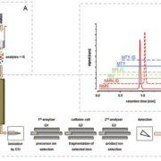
Progress in both LC-MS/MS and online sample preparation technology (pre-analytics) has advanced the analytical sensitivity of this methodology to the extent that for the measurement of many steroid hormones, modern MS applications have now transcended conventional IA methods in this regard. An example of this is the high sensitivity measurement of serum estradiol. External quality assurance data reveals that a wide range of concentrations can be obtained by immunoassay when measuring samples for estradiol at lower concentrations. Furthermore, a recent position statement from the Endocrine Society has stressed the need for better analytical methods to address the current poor performance of assays for measuring low concentrations of estradiol [4]. To this end, our group has developed a novel direct assay that is applicable to routine clinical use for the measurement of estradiol and estrone (therefore permitting calculation of total estrogen status) in male patients and patients on aromatase inhibitors [5]. This high sensitivity assay uses ammonium fluoride in the mobile phase to facilitate more efficient ionization and thereby increase analytical sensitivity. Additionally, an on-line solid phase extraction (OSM) system [Figure 2 (Waters, Manchester, UK)] allows a large volume of extract to be loaded and this coupled with a XEVO™TQS tandem mass spectrometer enables unprecedented analytical sensitivity to be achieved.
Conclusions and future prospects
LC-MS/MS is a very powerful tool which is enabling substantial innovations in the endocrine laboratory. Indeed, it is likely that the majority of emerging small molecules will be addressed by LC-MS/MS analysis. There are two keys areas in which future research and development for LC-MS/MS ought to be directed. Firstly, the utility of LC-MS/MS for the quantification of peptide hormones and proteins is already becoming a reality with published methods available for measurement of renin activity [6], parathyroid hormone [7] and insulin-like growth factor-1 [8] amongst others. These current methods require the skills of highly trained personnel in order to develop and run these assays, and it is hoped that continued innovation in this area will culminate in the development of rapid protein assays that are applicable to routine clinical use. Secondly, it seems feasible with existing technology to develop fully automated random-access LC-MS/MS analysers that will enable greater ease of use in non-specialist laboratory settings. However, the automation of mass spectrometry will not be achieved without a concerted effort from the in vitro diagnostics industry to fully realize the potential of LC-MS/MS across clinical medicine.
References
1. Monaghan PJ, Keevil BG, Trainer PJ. The use of mass spectrometry to improve the diagnosis and the management of the HPA axis. Rev Endocr Metab Disord 2013 Mar 15. [Epub ahead of print].
2. Monaghan PJ, Owen LJ, Trainer PJ, Brabant G, Keevil BG, Darby D. Comparison of serum cortisol measurement by immunoassay and liquid chromatography-tandem mass spectrometry in patients receiving the 11β-hydroxylase inhibitor metyrapone. Ann Clin Biochem 2011; 48: 441–446.
3. Monaghan PJ, Owen LJ, Trainer PJ, Brabant G, Keevil BG, Darby D. Response to ‘Comparison of serum cortisol measurement by immunoassay and liquid chromatography-tandem mass spectrometry in patients receiving the 11β-hydroxylase inhibitor metyrapone’ by Halsall DJ and Gurnell M. Ann Clin Biochem 2012; 49: 204–205.
4. Rosner W, et al. Challenges to the measurement of estradiol: An Endocrine Society Position Statement. J Clin Endocrinol Metab 2013; 98: 1376–1387.
5. Owen LJ, Wu FC, Labrie F, Keevil BG. A rapid direct assay for the routine measurement of oestradiol and oestrone by LC-MS/MS. Ann Clin Biochem [In press].
6. Carter S, Owen LJ, Kerstens MN, Dullaart RP, Keevil BG. A liquid chromatography tandem mass spectrometry assay for plasma renin activity using online solid-phase extraction. Ann Clin Biochem 2012; 49: 570–579.
7. Kumar V, Barnidge DR, Chen LS, Twentyman JM, Cradic KW, Grebe SK, Singh RJ. Quantification of serum 1-84 parathyroid hormone in patients with hyperparathyroidism by immunocapture in situ digestion liquid chromatography-tandem mass spectrometry. Clin Chem 2010; 56: 306–313.
8. Kay R, Halsall DJ, Annamalai AK, et al. A novel mass spectrometry-based method for determining insulin-like growth factor 1: assessment in a cohort of subjects with newly diagnosed acromegaly. Clin Endocrinol 2013; 78: 424–430.
9. Sturgeon CM, Viljoen A. Analytical error and interference in immunoassay: Minimizing risk. Ann Clin Biochem 2011; 48: 418–432.
10. Vogeser M, et al. Pitfalls associated with the use of liquid chromatography-tandem mass spectrometry in the clinical laboratory. Clin Chem 2010; 56: 1234–1244.
11. Duxbury K, Owen LJ, Gillingwater S, Keevil BG. Naturally occurring isotopes of an analyte can interfere with doubly deuterated internal standard measurement. Ann Clin Biochem 2008; 45: 210–212.
12. Davison AS, Milan AM, Dutton JJ. Potential problems with using deuterated internal standards for liquid chromatography-tandem mass spectrometry. Ann Clin Biochem 2013; 50: 274.
13. Twentyman JM, Cradic KW, Singh RJ, Grebe SK. Ionic cross talk can lead to overestimation of 3-methoxytyramine during quantification of metanephrines by mass spectrometry. Clin Chem 2012; 58: 1156–1158.
The authors
Phillip J. Monaghan*1 PhD, Laura J. Owen2 MSc, Peter J Trainer3 MD, and Brian G Keevil2 MSc
1Department of Clinical Biochemistry, 3Department of Endocrinology, The Christie NHS Foundation Trust, Wilmslow Road, Manchester M20 4BX, UK.
2Department of Clinical Biochemistry, University Hospital of South Manchester, Southmoor Road, Manchester, M23 9LT, UK.
*Corresponding author
E-mail: Phillip.Monaghan@nhs.net
The early diagnosis of common colds caused by coronavirus is a crucial step in preventing the recurrence of a global outbreak. The goals of this article are to discuss a prokaryotic-expressed recombinant nucleocapsid protein used in the development of a sensitive diagnostic assay for the diagnosis of human coronavirus infection.
by Dr Ming-Hon Hou
An overview of coronavirus
Human coronavirus (HCoV) was identified in the 1960s and has generally been associated with symptoms of the common cold. Although HCoV infections are generally mild, more severe upper and lower respiratory tract infections, such as bronchiolitis and pneumonia, which are particularly severe in infants, elderly individuals, and immunocompromised patients, have been documented. There have also been reports of clusters of HCoV infections that cause pneumonia in adults. In addition, a previous study reported that the neurotropism and neuroinvasion of HCoV are associated with multiple sclerosis.
In recent years, several emerging human coronaviruses have been discovered, and between 2003 and 2004, the SARS-CoV outbreak caused a worldwide epidemic that had a significant economic impact in countries where the disease outbreak occurred. Phylogenetic analyses have shown that SARS-CoV contains sequences that are closely related to sequences found in the betacoronaviruses. In 2004, another alphacoronavirus, HCoV-NL63, which was isolated from a 7-month-old child suffering from bronchiolitis and conjunctivitis, was reported in the Netherlands. In 2005, a novel betacoronavirus, HKU1 was found in patients with respiratory tract infections. Recently, a novel SARS-like coronavirus was found in patients with respiratory tract infections in the Middle East.
The RNA genomes of coronaviruses include genes encoding the structural proteins S (spike), M (matrix), E (envelope), and N (nucleocapsid). Additionally, some coronaviruses encode a third glycoprotein, HE (hemagglutinin-esterase), which is present in most of the betacoronaviruses. A helical nucleocapsid exists in the centre of the viral particle. The primary function of CoV N protein (NP) is to recognize a stretch of RNA that serves as a packaging signal, leading to the formation of the ribonucleoprotein (RNP) complex or to a long helical nucleocapsid structure during viral assembly. The formation of the RNP is important for maintaining the RNA in an ordered conformation suitable for replication and transcription of the viral genome. The CoV NP was shown to be involved in the regulation of cellular processes, such as gene transcription, actin reorganization, host cell cycle progression, and apoptosis.
Coronaviruses cause colds of mild to moderate severity and are transmitted by aerosols of respiratory secretions, the fecal–oral route, and mechanical transmission. The most common symptoms of coronavirus infection are nasal catarrh and a sore throat, and the illness typically lasts approximately 6 to 8 days. The early diagnosis of common colds caused by a coronavirus is an important step in preventing the recurrence of a global outbreak. Previously, rapid viral diagnosis has also been critical in the control of epidemics and the management of SARS patients. HCoVs are difficult to detect, and the current diagnosis of coronaviral infection is based on reverse transcription polymerase chain reaction with real-time PCR and antibody detection.
Previous studies have shown that NPs are the immunodominant domain in hosts infected with several coronaviruses. Additionally, it has been shown that NPs can accumulate intracellularly before being packaged into mature viruses and are the most abundant viral protein. NP is involved in the pathological reaction to human coronavirus and is a key antigen for the development of a sensitive diagnostic assay. These characteristics make NP a suitable candidate for the early diagnosis of coronavirus infection.
Nucleocapsid protein for coronavirus serodiagnosis
NP is involved in the pathological reaction to CoV infection and has been used in the development of a sensitive diagnostic assay. Previous studies reported that NP can be detected in the serum samples of SARS patients as early as 1 day after disease onset. Prokaryotic-expressed NPs have successfully been used as antigens for the detection of antibodies specific to many viruses, including SARS-CoV and several animal coronaviruses, and were produced for establishing an antigen-capture ELISA (or Western blot assay) for the diagnosis of HCoV infection
These methods are highly sensitive and specific. For example, Shi et al. [10] used recombinant SARS-CoV NP to establish an antigen-capture ELISA for SARS diagnosis. Anti-NP antibodies could be detected in approximately 90% of SARS patients 11 to 61 days after illness. No false positives were observed in non-SARS patients or health care workers.
An immunofluorescence assay is the gold standard for the detection of SARS. However, it requires efficient SARS-CoV replication in vitro to use whole virus or infected cell lysates as antigens. There are several reasons for selecting a recombinant protein rather than whole virus for this assay. The prokaryotic expression system is high yield, inexpensive, highly efficient, does not require viral cultures, and is non-toxic. Despite these advantages, viral proteins expressed in prokaryotic cells lack post-translational modifications that are present in proteins expressed in baculovirus expression systems.
Using recombinant nucleocapsid protein as an antigen for coronavirus infection diagnosis: one recent case study
HCoV is distributed worldwide. Recently, we produced soluble recombinant human coronavirus OC43 (HCoV-OC43) NP to analyse the antigenicity of the betacoronavirus HCoV-OC43 NP. To express soluble HCoV-OC43 NP as a set of fusion proteins in E. coli, the NP gene was cloned into the pET-28a expression vector. His-tagged NP was purified from the soluble fraction using Ni-NTA column chromatography [Figure 1]. The yield from 1 L of bacterial culture was as large as 10 mg of pure NP after extraction and column chromatography. A recombinant protein-based Western blot assay was used to screen human serum from young adults, middle-aged and elderly patients with respiratory infection symptoms and cord blood units.
Western blotting is generally accepted as the most effective method for unequivocally locating linear or continuous immunodominant epitopes. Pohl-Kooppe et al. [8] also reported that Western blotting is a more sensitive test system than an immunofluorescence assay for the analysis of sera from pediatric groups. Our results showed that approximately 80–90% of serum samples from young adults and middle-aged and elderly patients with respiratory infections reacted strongly to the HCoV-OC43 NP, indicating prior exposure to this disease. In addition, the serum samples tested in this study were 81% seropositive for HCo-229E NP [Fig. 2].
This finding is consistent with previous epidemiological surveys that concluded that seroprevalence increases rapidly during childhood, attaining a seroprevalence rate of up to 90% in adults. Additionally, antibodies against HCoV-OC43 NP were detected in over 90% of cord blood samples tested. Maternally acquired antibodies may help to protect a newborn baby from HCoV-OC43 infection, although this protection appears to wane by 4 to 5 months of age. HCoV is responsible for approximately 30% of all common colds, and it is expected that 80–90% of serum samples from healthy donors and patients have antibodies to HCoV-OC43.
CoV NPs contain multiple immunodominant epitopes and antigenic sites. To compare the immunoreactivity of the three structural regions of HCoV-OC43 NP, three truncated recombinant fragments [aa 1–173 (the N-terminal domain), aa 174–300 (the central region), and aa 301–448 (the C-terminal domain)] were produced in E. coli; these regions were chosen based on PONDR (predictor of naturally disordered regions) predictions. The reactivity of human serum against these fragments was determined through Western blotting. The human serum reacted strongly with the central region and the C-terminal domain of the NP, whereas the N-terminal domain demonstrated low reactivity with the antibody. The findings of the current study are consistent with those of Chen et al. [2], who found that the antigenicity of the C-terminus of SARS-CoV NP was higher than that of the N-terminus.
The polyclonal antibody against coronavirus NP could be used to develop a rapid, easy and specific diagnostic tool for the detection of HCoV infections through immunofluorescence or ELISA-based tests. Many studies have reported that NP polyclonal antibody does not cross-react with other human CoV NPs, including those of SARS-CoV and HCoV-229E, despite the presence of highly conserved motifs in these coronavirus NPs. Previous studies also showed that the anti-SARS CoV NP and anti-HCoV-229E NP polyclonal antibodies did not cross-react with other human CoV NPs.
In our recent studies, using purified recombinant NP as an antigen, a polyclonal antibody was generated from rabbit serum with specificity for HCoV-OC43 NP; this antibody reacted specifically with HCoV-OC43 NP and did not cross-react with other human CoV NPs (including those of SARS-CoV and 229E) through Western blotting.
Conclusion
A novel SARS-like coronavirus was found in patients with respiratory tract infections in the Middle East. Thus, new and convenient diagnostic methods for CoV infection are urgently needed. The prokaryotic expression of recombinant HCoV NP is suitable for high-sensitivity, highly specific antibody production and can be used for the epidemiological screening of HCoV infection in the future.
References
1. Che XY, Qiu LW, Liao ZY, Wang YD, Wen K, Pan YX, Hao W, Mei YB, Cheng VC, Yuen KY. Antigenic cross-reactivity between severe acute respiratory syndrome-associated coronavirus and human coronaviruses 229E and OC43. J Infect Dis 2005; 191: 2033–7.
2. Chen Z, Pei D, Jiang L, Song Y, Wang J, Wang H, Zhou D, Zhai J, Du Z, Li B, Qiu M, Han Y, Guo Z, Yang R. Antigenicity analysis of different regions of the severe acute respiratory syndrome coronavirus nucleocapsid protein. Clinical Chem 2004; 50: 988–95.
3. He Q, Chong KH, Chng HH, Leung B, Ling AE, Wei T, Chan SW, Ooi EE, Kwang J. Development of a Western blot assay for detection of antibodies against coronavirus causing severe acute respiratory syndrome. Clin Diagn Lab Immunol 2004; 114: 417–22.
4. Huang CY, Hsu YL, Chiang WL, Hou MH. Elucidation of the stability and functional regions of the human coronavirus OC43 nucleocapsid protein. Protein Sci 2009; 18: 2209–18.
5. Huang LR, Chiu CM, Yeh SH, Huang WH, Hsueh PR, Yang WZ, Yang JY, Su IJ, Chang SC, Chen PJ. Evaluation of antibody responses against SARS coronaviral nucleocapsid or spike proteins by immunoblotting or ELISA. Journal Med Virol 2004; 73: 338–46.
6. Liang FY, Lin LC, Ying TH, Yao CW, Tang TK, Chen YW, Hou MH. Immunoreactivity characterisation of the three structural regions of the human coronavirus OC43 nucleocapsid protein by Western blot: Implications for the diagnosis of coronavirus infection. J Virol Methods 2013; 187: 413–20.
7. Mourez T, Vabret A, Han Y, Dina J, Legrand L, Corbet S, Freymuth F. Baculovirus expression of HCoV-OC43 nucleocapsid protein and development of a Western blot assay for detection of human antibodies against HCoV-OC43. J Virol Methods 2007; 139: 175–80.
8. Pohl-Koppe A, Raabe T, Siddell SG, ter Meulen V. Detection of human coronavirus 229E-specific antibodies using recombinant fusion proteins. J Virol Methods 1995; 55: 175–83.
9. Shao X, Guo X, Esper F, Weibel C, Kahn JS. Seroepidemiology of group I human coronaviruses in children. J Clin Virol 2007; 40: 207–13.
10. Shi Y, Yi Y, Li P, Kuang T, Li L, Dong M, Ma Q, Cao C. Diagnosis of severe acute respiratory syndrome (SARS) by detection of SARS coronavirus nucleocapsid antibodies in an antigen-capturing enzyme-linked immunosorbent assay. J Clin Microbiol 2003; 41; 5781–2.
11. Timani KA, Ye L, Zhu Y, Wu Z, Gong Z. Cloning, sequencing, expression, and purification of SARS-associated coronavirus nucleocapsid protein for serodiagnosis of SARS. J Clin Virol 2004; 30: 309–12.
The author
Ming-Hon Hou PhD
1 Biotechnology Center, National Chung Hsing University, Taichung, Taiwan
2 College of Life Science, National Chung Hsing University, Taichung, Taiwan
3 Institute of Genomics and Bioinformatics, National Chung Hsing University,
Taichung, Taiwan
E-mail: mhho@dragon.nchu.edu.tw
Helicobacter pylori (Hp) -infection and atrophic gastritis (AG) are the most important risk conditions preceding gastric cancer (GC). Following extensive research and development, a Finnish biotechnology company, Biohit Oyj, has launched the GastroPanel test, a panel of four stomach-specific biomarkers that give accurate information on both the structure and function of gastric mucosa.
by Dr Kari Syrjänen
Since the risk of GC and peptic ulcer disease among individuals with healthy stomach is very low, it is essential to distinguish between subjects with healthy stomach and those with gastric disorders. With GastroPanel – a simple blood test – it is now possible to detect the patients who are at high risk for GC because they harbour either Hp -infection, AG or both in their stomach mucosa. Hp -infection alone increases the risk of GC several-fold, and this risk is over 90-fold among patients with Hp -related severe AG of both the corpus and antrum (pangastritis)[1, 3].
Another area of use for the GP test are the dyspeptic complaints, which in western countries appear in 20-40% of the population. According to most current medical practices, the assessment of these complaints should invariably include a gastroscopic examination for which the existing resources are clearly insufficient and which is actually not really necessary. The same applies to the costly and risky “test” medications with proton-pump inhibitors (PPIs), since it is now possible to screen the patients at true risk and for whom gastroscopy is indicated by using the GastroPanel test. With this approach, approximately 40-70% of the limited and expensive endoscopy capacity can be released for colonoscopies, i.e. for screening and early detection of colorectal cancer. Because of the fact that, particularly among the elderly, dyspeptic complaints are frequently of large intestinal origin, it is cost-effective to supplement the examinations of these dyspeptic patients with colorectal screening methods like the ColonView test, a stool based detection of Hb and Hb/Hp complex, or colonoscopy.
The safe and cost-effective GastroPanel test enables early detection of many different disorders, and thus helps avoiding the majority of subsequent health problems. Besides gastric and esophageal cancer, undetected AG of the corpus (acid-free stomach) can eventually also lead to malabsorption of vitamin B12, iron, magnesium, calcium, and some drugs. AG of the antrum, in turn, increases the risk of peptic ulcer disease and GC.
Concomitant AG of the antrum and corpus (pangastritis) is the single most important risk condition for GC. A minority of GCs can develop directly from HP-induced gastritis, without recognizable stages of mucosal atrophy. It is well known that vitamin B12 deficiency can lead to pernicious anemia (PA), dementia, depression, and injuries of the peripheral nervous system. Calcium deficiency, in turn, leads to osteoporosis. The absorption of many drugs is impaired in acid-free stomach. The risk of serious intestinal infections (giardiasis, malaria, Clostridium difficile and E. coli EHEC) can be increased particularly among senior citizens with AG.
Within public healthcare, it is possible to achieve substantial cost savings by replacing the systematic use of gastroscopy with a simple and inexpensive first-line diagnostic tool like the GastroPanel test for all patients with dyspeptic symptoms.
References
1. Suovaniemi O. GastroPanel-tutkimus osaksi dyspepsian hoitokäytäntöä. Yleislääkäri 2007; 4:104-106.
2. Malfertheiner P, Mégraud F, O’Morain C ym. Current concepts in the management of Helicobacter pylori infection: the Maastricht III Consensus Report. Gut 2007; 56:772-781.
3. Agreus L, Kuipers EJ, Kupcinskas L, Malfertheiner P, Di Mario F, Leja M, Mahachai V, Yaron N, van Oijen M, Perez Perez G, Rugge M, Ronkainen J, Salaspuro M, Sipponen P, Sugano K, Sung J. Rationale in diagnosis and screening of atrophic gastritis with stomach-specific plasma biomarkers. Scand J Gastroenterol 2012; 47:136-147.
The authors
Prof Kari Syrjänen,* MD, PhD, FIAC,
Chief Medical Director, Biohit Oyj.
Lea Paloheimo PhD
Director of Business Development and Quality, EurClinChem,
*Corresponding author
E-mail: kari.syrjanen@biohit.fi
Mass spectrometry is poised for a new era, as clinical labs and researchers, hospital managers and industry prepare themselves for expansion in its use. Fuelling growth are trends towards personalized healthcare, the identification of novel biomarkers for translational medicine, large-scale epidemiological screenings as well as everyday clinical chemistry tests beyond just toxicology and endocrinology. There is room for such growth. At present, clinical lab applications of mass spectrometers account for only about 5% of the market.
Superior sensitivity and specificity, samples reusable
Mass spectrometry identifies a molecule by its unique mass-to-charge ratio, and is both highly sensitive and specific. In spite of concerns about cost and steep learning curves, the superiority of mass spectrometry versus immunoassays has never been disputed. Indeed, a study by the US National Cancer Institute (NCI) in 2008 focused on using mass spectrometry to distinguish between breath samples from patients with ovarian epithelial cancer versus those with polycystic ovarian syndrome or endometriosis.
Another advantage of mass spectrometry is its ability to use the same serum for multiple analyte profiling. This makes it useful in large-scale clinical studies, where samples have often been archived. Another NCI study, for instance, used mass spectrometry to identify biomarkers in blood from patients with acute myeloid leukemia; some of the samples were almost 10 years old. Dated samples have also been used for a range of other biomarkers, including malignant melanoma, soft tissue sarcomas and non-small cell lung cancer.
Gas chromatography and liquid chromatography
As a technology, mass spectrometry is not new in a lab setting. Gas chromatograph MS (GC-MS) has been used for ages in the diagnosis of organic metabolic disorders. More recently, liquid chromatograph mass spectrometry (LC-MS) has become a recommended resource for screening newborns.
The longer use of GC-MS means a bigger user base, as well as a more extensive legacy database, richer software libraries and advanced algorithms. Although GC-MS requires more complex processes for sample preparation (discussed below), it is relatively inexpensive compared to LC-MS systems, and has been considered effective enough for the bulk of applications.
The challenge of standardization
However, there is still some way to go before mass spectrometry attains wider use. One key barrier is a lack of standardization, above all in the preparation of samples. Clinical labs have different approaches to this issue, especially in terms of purification. This leads to sometimes-significant differences in results. Confounding the problem are continuing changes in the methods used for sample preparation, over time even within individual laboratories.
In the US, the Clinical and Laboratory Standards Institute has published two sets of recommendations on the use of MS. However, these leave quite a bit of room for interpretation and are considered no more than broad guidelines.
Preparation of samples for mass spectrometry
Typically, two steps are involved in preparing a sample: the concentrating of analytes, followed by ionization. The sample itself consists of two parts: the analytes of interest, and other components which are collectively known as the sample matrix. Sample preparation is considered the most difficult when whole blood or fractions are involved, given a relatively low density of analytes. Urine lies at the the other end of the spectrum, since the kidneys have already done most of the job of concentrating analytes.
Techniques for preparing samples include solid-phase extraction (SPE), immunoextraction (or immunoaffinity purification) and so-called ‘dilute-and- shoot’. In SPE, analytes and other matrix compounds are separated on the basis of their physical and chemical properties, among them charge and polarity. SPE systems consist of a liquid, mobile phase and a solid stationary phase (usually disposable cartridge-based). The liquid phase uses two different solvents, one for binding and washing, and another for elution.
Immunoextraction separates antibodies bound to the analytes from ‘free’ matrix components, by immobilizing them to a chromatographic column or polystyrene beads. After incubation with an immobilized antibody, unwanted components are washed away, and the enriched analyte is then eluted; another method is to concentrate the sample by drying, followed by re-suspension and injection into the chromatography system.
The third mechanism for preparing MS samples, dilute-and-shoot, is generally used in samples with a relatively high concentrations of analytes (e.g. urine). Here, dilution is usually effective enough to reduce matrix components to a
manageable level.
Successful ionization essential
The process of analysis relies wholly on successful ionization, as mass spectrometers can only detect charged analytes in a gaseous phase. Ionization can be either positive (cationic) or negative (anionic). The most common techniques for ionization in a clinical lab consist of chemical ionization and electrospray.
Chemical ionization generates ions by combining heat and plasma (produced by high-voltage electricity), at atmospheric pressure. While high temperatures vaporize the sample, the plasma (also known as a corona discharge) ionizes the evaporated solvent. Following this, mechanical interaction of the sample components (including analytes of interest) leads to the formation of negative or positive ions.
On its part, electrospray ionization uses electricity, heat and air to successively reduce the size of droplets that elute off the chromatographic column and sharply increase their charge. Ions (above all, proteins) desorb from the liquid droplet surface into a gas phase and then enter the mass spectrometer.
Challenges for vendors
Until recently, industry has focused on process improvements, while researchers have concentrated on improving the specificity and sensitivity of mass spectroscopy. Innovations from vendors have aimed at increasing the efficiency of ionization and of ion transfer, and accelerating discovery of biomarkers by combining size exclusion and affinity capture to enrich low molecular weight proteins, and more quickly separate diseased from clear samples. Some companies have also coupled reference databases of micro-organisms to their mass spectroscopy systems.
The greatest challenge for industry, however, has been to increase user acceptability. Research scientists rather than clinical lab technologists have been the traditional target for mass spectrometry manufacturers. The former, typically, have more interest in top-of-the-line technical specifications and performance than user-friendliness. The potential demand from clinical labs is forcing vendors to change approach. As a result, several are now beginning to package equipment sales with training and support.
Industry is also paying attention to systems integration, to bundle sample preparation instrumentation into a mass spectrometry suite and control its findings. Indeed, software has so far proved to be one of the biggest impediments to the growth of mass spectrometry, once again given the delicate balance between enabling new users to operate a system on the one side, while permitting complex adjustment of performance parameters on the other. OEMs have sought to plug this gap with bespoke add-ons but, as all IT systems designers know, this adds to system cost.
Researchers aim for more precision, ease of use
On the R&D side, a potentially promising area consists of so-called time-of-flight (TOF) mass spectrometers. TOF provides accuracy of 1 part per million by accelerating gas phase ions toward a detector via an electric field. Other initiatives are focused on robotic assistance, turbulent-flow chromatography and ion mobility – with considerable potential seen in linear ion traps. Scientists are also exploring the use of nanospray interfaces as well as microfluidics, though most successes to date have been at bench scale. In the future, such improvements will permit a reduction of detection thresholds, along with greater precision, ease of use and efficiency.
Some trade-offs inevitable
For both researchers and industry, the Holy Grail is to devise adequate user-configurability for trade-offs between high throughput on one side (required, for example, in epidemiological studies or newborn screening), and sensitivity and specificity on the other. Even now, detection of steroids such as cortisol, estradiol and testosterone remain a challenge at the lower end of their reference range, but require high precision in certain categories of patients, for example elderly female patients.
Lab use of mass spectrometry still minor, room for growth
No one doubts that the market for mass spectrometry is potentially huge. Globally, sales have been rising briskly, after falling due to the recession. A study from Los Angeles-based Strategic Directions International estimates the 2011 mass spectrometer market at USD 3.9 billion, with projections of USD 4.8 billion by 2014. The US and Canada hold the largest share of the market (38%) followed by Europe (31%) and Japan (13%), with other countries accounting for the remainder. Leaders in the mass spectrometer market include AB Sciex, Thermo Fisher Scientific, Waters and Agilent Technologies (all from the US), along with Hitachi and Shimadzu. European companies have a smaller presence, and include Germany’s GSG, Spectromat and Thermolinear.
As mentioned before, the clinical lab segment accounts for a very small share of total sales. The biggest users are pharmaceutical companies (a share of 20% of sales, with mass spectrometers increasingly used for metabolomic screening and drug discovery). Government follows closely (with an 18.5% share), universities (12.6%) and environmental/general testing services (9.4%). Electronics, the food and chemical industries also buy more mass spectrometers than clinical laboratories or hospitals.
However, the hope is that continuing growth in this entrenched base of other users will drive down unit costs of mass spectrometers, just as clinical labs get ready to increase their own requirements.
November 2024
The leading international magazine for Clinical laboratory Equipment for everyone in the Vitro diagnostics
Beukenlaan 137
5616 VD Eindhoven
The Netherlands
+31 85064 55 82
info@clinlabint.com
PanGlobal Media is not responsible for any error or omission that might occur in the electronic display of product or company data.
This site uses cookies. By continuing to browse the site, you are agreeing to our use of cookies.
Accept settingsHide notification onlyCookie settingsWe may ask you to place cookies on your device. We use cookies to let us know when you visit our websites, how you interact with us, to enrich your user experience and to customise your relationship with our website.
Click on the different sections for more information. You can also change some of your preferences. Please note that blocking some types of cookies may affect your experience on our websites and the services we can provide.
These cookies are strictly necessary to provide you with services available through our website and to use some of its features.
Because these cookies are strictly necessary to provide the website, refusing them will affect the functioning of our site. You can always block or delete cookies by changing your browser settings and block all cookies on this website forcibly. But this will always ask you to accept/refuse cookies when you visit our site again.
We fully respect if you want to refuse cookies, but to avoid asking you each time again to kindly allow us to store a cookie for that purpose. You are always free to unsubscribe or other cookies to get a better experience. If you refuse cookies, we will delete all cookies set in our domain.
We provide you with a list of cookies stored on your computer in our domain, so that you can check what we have stored. For security reasons, we cannot display or modify cookies from other domains. You can check these in your browser's security settings.
.These cookies collect information that is used in aggregate form to help us understand how our website is used or how effective our marketing campaigns are, or to help us customise our website and application for you to improve your experience.
If you do not want us to track your visit to our site, you can disable this in your browser here:
.
We also use various external services such as Google Webfonts, Google Maps and external video providers. Since these providers may collect personal data such as your IP address, you can block them here. Please note that this may significantly reduce the functionality and appearance of our site. Changes will only be effective once you reload the page
Google Webfont Settings:
Google Maps Settings:
Google reCaptcha settings:
Vimeo and Youtube videos embedding:
.U kunt meer lezen over onze cookies en privacy-instellingen op onze Privacybeleid-pagina.
Privacy policy