Beukenlaan 137
5616 VD Eindhoven
The Netherlands
+31 85064 55 82
info@clinlabint.com
PanGlobal Media is not responsible for any error or omission that might occur in the electronic display of product or company data.
Worldwide, screening has been shown to reduce mortality and incidence of colorectal cancer. Despite its documented success, people still fail to participate and screening rates remain low in most countries. Given that patient-reported barriers include resistance to recommended fecal-based methods or endoscopy, blood-based tests have the potential to increase participation in colorectal cancer screening programmes.
by Dr Theo deVos
Background
Globally, colorectal cancer (CRC) is the third most common cancer in men and the second in women, with an estimated 1.36 million cases and causing an estimated 694,000 deaths in 2012 [1]. These rates are unnecessarily high since CRC is an excellent candidate for screening as evidenced by large randomized trials demonstrating reductions in mortality and incidence [reviewed in 2, 3]. Biologically, CRC usually develops slowly, going through a progression from non-cancerous polyp to cancer over a period of a decade or more. This biology readily lends itself to screening and early detection which has a significant positive impact on the effectiveness of intervention. For example, in the United States, 5-year survival is ~90 % if the tumour is confined locally when detected, ~70% if it has spread regionally, but only ~10% if distant metastases are present [4].
Colonoscopy is the predominantly recommended method for routine screening in some countries including the United States, as it enables detection and intervention in the same procedure. It is also the diagnostic follow-up for positive results of other screening tests. However, challenges with capacity and quality, financial concerns, and patient resistance have led to its lack of use as the primary screening modality in most settings. In some countries, flexible sigmoidoscopy is showing a resurgence, with reports demonstrating mortality and incidence benefits [2]. Table 1 displays a list of common CRC screening methods along with new methods coming on-line, today.
The first non-invasive tests for CRC were based on the detection of fecal occult blood (FOBT), and these have been further developed into immunological tests (FIT) using specific antibodies to detect hemoglobin. These tests are typically designed to allow patients to collect stool samples at home and ship the sample by mail to a central laboratory for testing. A newer alternative to fecal blood testing is the analysis of genetic/epigenetic markers in fecal material. This is the basis for the Cologuard test (Exact Sciences, WI, USA), a fecal DNA test recently approved by the US FDA [5]. Blood-based screening tests that measure tumour biomarkers in plasma or serum have been developed as a minimally-invasive alternative to fecal testing. DNA methylation tests based on SEPT9 have become available in Europe and are undergoing regulatory review in China. In addition, methylated SEPT9 testing is available as laboratory-developed tests (LDTs) in the USA, and a kitted version (Epi proColon®; Epigenomics AG, Germany) is currently undergoing US FDA premarket (PMA) review [6]. Another blood-based test, the ColonSentry risk test based on an expression panel is available as an LDT in the USA and in Japan.
Given the clear benefit of screening and the long standing availability of tests, the lack of participation is disappointing, and improving screening rates is a broadly accepted goal. As an example, the ‘80 by 2018’ campaign in the USA has set a goal of 80% adherence to screening guidelines by 2018 [7]. In order to meet this goal, barriers that prevent screening must be understood and overcome. There are numerous reports focused on understanding patient barriers to CRC screening. Although this is a complex issue involving costs, time, physician recommendation and several other factors, one consistent message from these studies is that the test methods themselves present barriers. Many patients are uncomfortable with all or part of the colonoscopy process and many are also uncomfortable with collecting and shipping fecal samples [8]. As a consequence, CRCs are diagnosed symptomatically in more instances than necessary, when the disease has spread beyond the primary site, resulting in greatly reduced survival rates. The availability of a screening test using a simple and common blood draw, which can be included as part of a regular check-up, has the potential to overcome some barriers and improve screening rates.
Blood-based screening
There are a number of approaches to the measurement of cancer biomarkers in the blood. The detection and quantification of circulating tumour cells represents an early approach, which was developed into a commercial system (e.g. CellSearch; Janssen Diagnostics, NJ, USA) though this analysis has not generally been used for cancer screening. Another alternative derives from the isolation and fractionation of circulating immune cells and the quantification of gene expression panels correlated with the disease by reverse-transcriptase PCR. This ‘sentinel concept’ is the basis for the ColonSentry test (GeneNews, Canada) in Table 1. A third alternative is the measurement of metabolic products by mass-spectrometry that are correlated with the presence of cancer. As an example, a commercial test (Cologic; Phenomenome, Canada) was developed based on the measurement of serum levels of GTA-446, an anti-inflammatory fatty acid. The most developed and perhaps simplest approach in this field is the measurement of cell-free genetic or epigenetic markers in plasma or serum that are highly correlated with the presence of cancer. As shown in Table 1, the methylated Septin9 biomarker and the Epi proColon® test were developed based on this approach.
Screening biomarkers in plasma and serum
The recognition that tumour DNA contains genetic and epigenetic changes that can serve as biomarkers dates back a number of decades. As reviewed recently, the list of biomarker reports for colorectal cancer grows ever longer [9]. Although numerous studies report on marker performance, the majority of studies include only a limited number of cases and controls, and only a small subset of markers have been rigorously tested in the clinical setting. Furthermore, a review of marker studies in ClinicalTrials.gov indicated very few ongoing CRC marker screening trials. Well validated markers include methylated SEPT9 described above, and the methylation of BCAT1 and IKZF1 sequences in plasma which have shown to be correlated with CRC [10] and are currently being tested in a clinical trial in Australia. There are many interesting genetic and epigenetic markers, but most await additional validation data that will support clinical utility.
Laboratory considerations for a plasma-based screening test
The basic concept outlined in Figure 1 illustrates key points associated with development of a genetic/epigenetic screening test. CRC screening from blood samples imposes rigorous demands that impact the reduction to practice for a test including: (a) high volume (millions of tests); (b) low target copy number (~1 copy per mL); (c) fragmented DNA; (d) large sample size (e.g. 3.5 mL); and (e) kitted reagents. These are discussed using the methylated Septin9 test as a case study.
Blood draw and processing
Given that screening is a high volume activity, an inexpensive and standard sample collection method is beneficial. In this case, a simple blood draw using a standard collection tube (e.g. K2EDTA plasma collection tube) is performed at the clinic or draw station. Plasma or serum is separated and if necessary they can be re-centrifuged to ensure cell-free status. The emphasis is on preparing cell-free material to limit background contamination due to lysis of nucleated cells in the blood. While this has led to the use of specialized collections tubes (Streck, NE, USA) in the field of prenatal diagnostics, these have not been widely tested for colorectal cancer screening. Cleared plasma can be tested immediately, or stored frozen for a period of time.
Nucleic acid extraction
In this step, cell-free nucleic acids are extracted from the plasma sample. While a number of commercial methods have been developed for this purpose, it remains the Achilles heel of the process. Given the wide range in target concentration, and particularly the exceptionally low copy number expected for early cancers (in the single copy per mL range) [6], as well as the fragmented nature of cell-free DNA, the extraction methods must be designed to handle large samples (e.g. 3–4 mL of plasma), and be able to isolate fragmented DNA. The use of magnetic particles for purification coupled with modified binding and wash buffers designed to capture the full range of DNA fragments has simplified the extraction, and with the development of liquid handling platforms that can process larger volumes, this step is becoming automatable. While the reduction from 3.5 mL plasma to 100 µL of DNA eluate would raise concerns for PCR inhibition, for DNA methylation tests, it is possible to reduce the wash steps because the DNA is extensively purified in the bisulfite treatment process.
Bisulfite treatment
The bisulfite treatment process is required if the target is DNA methylation-based. Recent improvements in bisulfite conversion technology have simplified the treatment. The change to ammonium bisulfite allows for liquid reagents – a key attribute for kit development. In combination with elevated temperatures, bisulfite incubation time is reduced to less than 1 hour, enabling single shift turn-around times for tests. Furthermore, the reaction can be purified using a magnetic particle extraction that takes advantage of the same particles used for the initial DNA extraction. This process can also be automated on a standard liquid handling platform to improve throughput and quality.
Real-time PCR
For genetic (mutation)-based tests, the test can be performed immediately following initial DNA extraction, though it is important to increase the stringency of DNA washes to limit the potential for PCR inhibition. In the final steps, either genetic or epigenetic markers are measured by real-time PCR. For screening applications, the target concentration dictates the conditions and interpretation of the PCR reaction. For example, in the methylated Septin9 test, the final recovered bisulfite converted template DNA is split into three wells and run in three PCR reactions. Although the PCR reaction is run as a real-time assay, the test is essentially a qualitative end point test, since a well is called positive if a PCR curve occurs at any cycle during the course of the reaction. In addition, the results of the three reactions are combined to produce a final interpretation for a patient sample. For the CE-marked Epi proColon 2.0 product, the sample is called positive if two of three wells are positive. For the Ep proColon product undergoing US FDA PMA review, the sample is called positive if any of three wells are positive. This allows for a greater emphasis on a specific test parameter – for sensitivity (any well-positive) or test specificity (two out of three wells positive).
Summary
The use of genetic and epigenetic biomarkers for cancer screening is a field still in its infancy that has great opportunities for growth. Because these biomarkers can be used as indicators of disease, they also have diagnostic and prognostic potential that will be incorporated into the clinical-decision making process. For CRC screening, test kits are already available in Europe and other countries, and are currently under review by both the US and Chinese FDA organizations. In the US, LDTs are currently marketed, and together, all progress represents significant opportunities to generate positive momentum. The introduction of simple, blood-based screening would provide a viable alternative to patients refusing or avoiding current well established methods. The convenience factors of sample collection and processing by health professionals also avoids the challenges of faulty sampling, handling, and mailing associated with at-home self-collected tests. Finally, given the extensive collection of promising biomarkers on the horizon, mechanisms are needed now to expedite clinical utilization and validation to drive further improvements in test performance.
References
1. Ferlay J, Soerjomataram I, Ervik M, Dikshit R, Eser S, Mathers C, Rebelo M, Parkin DM, Forman D, Bray, F. GLOBOCAN 2012 v1.0, Cancer Incidence and Mortality Worldwide: IARC CancerBase No. 11 [Internet]. Lyon, France: International Agency for Research on Cancer; 2013. Available from: http://globocan.iarc.fr, accessed on 12/09/2014.
2. Kuipers EJ, Rösch T, Bretthauer M. Colorectal cancer screening – optimizing current strategies and new directions. Nat Rev Clin Oncol. 2013; 10: 130–142.
3. Brenner H, Stock C, Hoffmeister M. Effect of screening sigmoidoscopy and screening colonoscopy on colorectal cancer incidence and mortality: systematic review and meta-analysis of randomised controlled trials and observational studies. BMJ 2014; 348: g2467.
4. American Cancer Society. Colorectal Cancer Facts & Figures 2014-2016. Atlanta: American Cancer Society, 2014.
5. Imperiale TF, Ransohoff DF, Itzkowitz SH, Levin TR, Lavin P, Lidgard GP, Ahlquist DA, Berger BM. Multitarget stool DNA testing for colorectal-cancer screening. N Engl J Med. 2014; 370(14): 1287–1297.
6. Potter NT, Hurban P, White MN, Whitlock KD, Lofton-Day CE, Tetzner R, Koenig T, Quigley NB, Weiss G. Validation of a real-time PCR-based qualitative assay for the detection of methylated SEPT9 DNA in human plasma. Clin Chem. 2014; 60(9): 1183–1191.
7. National Colorectal Cancer Round Table. Tools & Resources – 80% by 2018. http://nccrt.org/about/80-percent-by-2018/
8. Gimeno García AZ. Factors influencing colorectal cancer screening participation. Gastroenterol Res Pract. 2012; 2012: 483417.
9. Toiyama Y, Okugawa Y, Goel A. DNA methylation and microRNA biomarkers for noninvasive detection of gastric and colorectal cancer. Biochem Biophys Res Commun. 2014; doi: 10.1016/j.bbrc.2014.08.001.
10. Mitchell SM, Ross JP, Drew HR, Ho T, Brown GS, Saunders NF, Duesing KR, Buckley MJ, Dunne R, Beetson I, Rand KN, McEvoy A, Thomas ML, Baker RT, Wattchow DA, Young GP, Lockett TJ, Pedersen SK, Lapointe LC, Molloy PL. A panel of genes methylated with high frequency in colorectal cancer. BMC Cancer 2014; 14: 54.
The author
Theo deVos PhD
Epigenomics Inc.,
Seattle, WA 98107, USA
E-mail: theo.devos@epigenomics.com
The state of Georgia is committed to supporting and growing the booming life science industry. By continually investing in its people, resources and solutions that meet companies’ unique business needs, Georgia is becoming a leader in the hematology and immunology sector.
When it comes to the study of blood and curing blood-related diseases, access to the right research, qualified talent and effective cold chain logistics is imperative to solving tomorrow’s health challenges.
These are a few of the many reasons Baxter International’s $1.3 billion (€0.96 billion) plasma fractionation plant, the America Red Cross’ Biomedical Services, the second largest blood processing facility in the world by volume, and Qualtex Laboratories, the United States’ largest independent testing lab for blood and plasma products, have chosen Georgia to call home.
Georgia is well equipped to support hematology companies and advanced research initiatives in treating and curing serious illnesses. In fact, Georgia’s robust hematology sector accounts for 21 percent of the state’s life science workforce.
Talent and training
One of the vital resources that companies in the hematology sector are finding in Georgia is access to the most innovative research and development, as well as the best and brightest talent. Georgia life science companies continue to take advantage of our world-class institutions and universities and the number one workforce training program in the nation, QuickStart.
In fact, EMSI ranks Georgia medical and health-related degrees as the No. 1 area of study in the region, and Georgia Tech’s biomedical/bioengineering programs are in the top five in the nation.
Emory University (Emory) has long been recognized for its expertise in cellular immunity and immune memory at the Emory Vaccine Center (EVC). EVC is one the largest and most comprehensive academic vaccine centres in the world. Nine in 10 HIV patients undergo lifesaving therapy that includes drugs that were created at Emory University.
Public-private partnerships
By connecting state resources such as the Centers for Disease Control and Prevention with companies and universities engaged in life science exploration, Georgia has created a dynamic and collaborative environment to support this booming industry.
The Atlanta Clinical & Translation Science Institute (ACTSI), a collaboration between Emory, the Georgia Institute of Technology (Tech) and the Morehouse School of Medicine, is solely focused on transforming scientific discovery into a positive impact on the community.
The Lam Lab, located at Emory and Tech, offers the ideal environment to foster integration between microtechnology development and experimental hematology and oncology practices.
The Georgia BioScience Training Center, a 50,000-square-foot facility dedicated to supporting research and technology transfer in the bioscience industry, will provide comprehensive, customized workforce training critical to the successful operations of bioscience and biomanufacturing industries. The Center will help train employees for Baxter’s $1 billion biomaufacturing plant in Covington, Ga.
Cold chain logistics
Supplying life-saving vaccines and treatments to the world would not be possible without Georgia’s extensive cold chain network. Being home to 75 facilities that have temperature-controlled and frozen storage capabilities, Georgia has built a strong value chain and an interconnected infrastructure to better meet the demands of the hematology sector. The Hartsfield-Jackson Atlanta International Airport houses a 32,000-square-foot Atlanta Perishables Center. The center is the only facility in the Southeast approved by the USDA to apply cold treatment, an alternative to methyl bromide.
Georgia Department of Economic Development www.georgia.org/hematology
Apitope, the drug discovery and development company focused on disease-modifying treatments for patients with autoimmune and allergic diseases, announced today that Bristol University research led by Apitope Founder and CSO, Prof David Wraith, on its treatment approach to autoimmune diseases, such as Multiple Sclerosis (MS), has been published in Nature Communications.
The researchers at the University of Bristol reported an important breakthrough in the fight against debilitating autoimmune diseases such as Multiple Sclerosis. Rather than the body’s immune system destroying its own tissue by mistake, researchers have discovered how cells convert from being aggressive to actually protecting against disease. It is hoped this latest insight will lead to the widespread use of antigen-specific immunotherapy as a treatment for many autoimmune disorders, including Multiple Sclerosis (MS), Factor VIII intolerance in hemophiliacs, Graves’ disease (hyperthyroidism) and uveitis, conditions for which Apitope is developing important new therapies.
Commenting on the research, Dr. Keith Martin, CEO said: “Multiple Sclerosis affects around 100,000 people in the UK and 2.5 million people worldwide. This is an important breakthrough in our fight against debilitating autoimmune diseases by providing further important information on how to stop cells attacking healthy body tissue. This research further reinforces Apitope’s treatment approach, which has already successfully completed two clinical trials in MS patients with MRI data showing a significant decrease in new lesions, and has the potential to improve the lives of millions of people worldwide. Importantly, we are now taking this approach into other serious autoimmune conditions as well as MS.”
The reported study, funded by the Wellcome Trust, is published in Nature Communications. The article entitled “Sequential transcriptional changes dictate safe and effective antigen-specific immunotherapy” that describes how researchers have managed to “switch off” autoimmune disease as a breakthrough for Multiple Sclerosis (MS) treatment, can be viewed on the following link: http://tinyurl.com/nupw4lj Apitope International NV, based in Belgium and the UK, is a drug developer of immunotherapies for the treatment of autoimmune and allergic diseases, including multiple sclerosis, factor VIII intolerance, uveitis and Graves’ disease. The company has a patented discovery platform which enables selection of potential disease-modifying peptide therapies for the autoimmune/allergic disease of interest; and has already generated a pipeline of 7 programmes in clinical and preclinical development, of which the lead programme in multiple sclerosis is partnered with Merck Serono.
Although known to the ancient Greeks, celiac disease was definitively demonstrated only in the late 1950s after development of the endoscope. In 1969, the new European Society for Pediatric Gastroenterology, Hepatology and Nutrition (ESPGHAN) codified the first diagnostic criteria for the disease.
Fast-growing challenge
The prevalence of celiac disease has exploded in recent years, above all in North America, Europe and Australia. The Association of European Coeliac Societies (AOECS) notes that those with the condition, but unaware of it, are compelled to live a life “filled with chronic pain and discomfort.”
If unchecked, inflammation caused by celiac disease seriously damages the lining of the small intestine, which produces enzymes for the digestion and absorption of food and essential nutrients. The malabsorption leads to diarrhea, weight loss and fatigue. Celiac disease eventually impacts on the bones, liver, brain and nervous system, in some cases seriously. AOECS includes “infertility, osteoporosis and small bowel cancer” in its list of long-term risk factors.
The role of prolamins
The principal protein responsible for celiac disease consists of prolamins, which are resistant to proteases and peptidases of the gut. They stimulate intestinal membrane cells in susceptible people to become permeable (or ‘leak’), by allowing larger peptides to bypass the sealant between cells, and thereby enter circulation.
The best understood prolamin is gliadin (in wheat). Other prolamins believed to play a role include hordein in barley, scelain in rye and zein in corn. The role of avenin in oats as a causative factor for celiac disease remains unclear. In Europe, however, the EU Commission requires that gluten-free oats are specially produced or processed to avoid contamination by wheat, rye and barley.
Prevalence growth ‘mystifying’, increase uneven
Estimates on the prevalence of celiac disease have leaped dramatically in recent years. It was previously believed that it affected about 1 in 1,500 people. However, new studies suggest a 15-fold higher rate, about 1 in 100 (1%) in both Europe, and the US. As the ‘New York Times’ observed last year, the spike in US prevalence of celiac disease is “mystifying.”
In Europe, prevalence varies widely. In the 30–64 year age group, the rate in Finland is eight times higher than in Germany (2.4% versus 0.3%). In addition, Finland has also shown a doubling in prevalence over 20 years – a fact which “cannot be explained by better detection rates.”
There is a higher prevalence of celiac disease in people with other conditions, such as Type 1 diabetes, Down Syndrome as well as both hypo- and hyper-thyroidism.
Genetics and environment
The challenges of celiac disease are manifold.
Its etiology is unclear. The disease is caused by “a combination of immunological responses to an environmental factor (gluten) and genetic factors.” The latter consist of the cellular receptors for two versions of HLA (human leukocyte antigen), DQ2 or DQ8. The absence of either results in “a negative predictive value … close to 100%.” This explains why people of Chinese, Japanese and African descent – who lack the HLA allele – are rarely diagnosed with celiac disease, unlike Caucasians.
Nevertheless, in a confirmation of the role of environmental triggers, the presence of HLA-DQ2 or -DQ8 is “necessary but not sufficient to predispose people to celiac disease.” In addition, the genes may be transmitted to some family members, but not others. First- and second-degree relatives of people with celiac disease show prevalence rates of about four-and-a-half and two-and-a-half times that of the general population.
The no-man’s land of gluten sensitivity
The symptoms of celiac disease are also varied, since it affects people differently. One of the best illustrations of the scale of the diagnostic challenge is the University of Chicago’s Celiac Disease Center, which lists as many as 300 symptoms that may accompany the disease.
Celiac disease is also routinely confused with irritable bowel syndrome. Indeed, a paper in 2009 published in the ‘American Journal of Gastroenterology’ remarks about the ‘no-man’s land of gluten sensitivity’ lying between celiac disease and irritable bowel syndrome.
As the US-based Celiac Disease Foundation observes, such factors make the disease difficult to diagnose. In addition, some patients have no symptoms at all.
Europe’s AOECS notes that only about 12%-15% of celiac disease patients obtain a diagnosis. In many cases, moreover, the time between experiencing first symptoms and diagnosis is over 10 years.
Confounding matters further is a lack of physician awareness about the onset of symptoms. Surveys in the US have shown that only 35% of primary care physicians had ever diagnosed celiac disease.
Peaks in diagnosis occur in childhood and between the fifth and seventh decades of life. The female-to-male ratio in celiac disease is about 2:1.
Strict diet only answer
There is no cure for celiac disease.
A gluten-free diet is used to manage symptoms and promote intestinal healing. The diet is strict and demanding. Patients can relapse if gluten is reintroduced, for some even in trace quantities, and people preparing gluten-free meals are urged to do so separately from other foods.
The only recommended preventative action against celiac disease is to avoid wheat-containing foods in an infant’s diet for six months after birth. Gluten increases the risk of developing celiac disease by five times, “within the first 3 months or after 7 months” of age.
In Europe, a 2006 EU Commission Directive bans the use of gluten containing foods in infant formula. The US, however, has no similar rule and the Celiac Disease Center at the University of Chicago simply notes that “most baby formulas are gluten-free.”
Guidelines for celiac disease
The original 1969 diagnostic criteria for CD by the European Society for Pediatric Gastroenterology, Hepatology and Nutrition (ESPGHAN) were revised in 1990, and most recently in 2011. Along with clinical guidelines from the North American Society for Pediatric Gastroenterology, Hepatology and Nutrition, these reflect the current consensus for celiac disease in pediatric practice.
In adults, testing for celiac disease is recommended only for symptomatic individuals and those in high risk groups. Screening is explicitly ruled out in asymptomatic patients, for example by the American Association of Family Physicians (AAFP).
Health authorities in many countries follow guidelines from the World Gastroenterology Organization (WGO).
For the WGO, celiac disease is based on patients having “characteristic histopathologic changes in an intestinal biopsy,” along with clinical improvement after a gluten-free diet.
The WGO’s latest guidelines specify serological tests for identifying patients in whom biopsy might be warranted, and investigating high-risk patients (including first- and second-degree relatives). The tests include immunoglobulin A (IgA) endomysial antibody (EMA), IgA anti-tissue transglutaminase antibody (tTG) and IgA and immunoglobulin G (IgG) deamidated gliadin peptide (DGP) antibodies. Small-bowel biopsy, however, is considered a ‘gold standard’ by the WGO.
The UK was one of the first countries to consider diagnosis and management of celiac disease in general practice. In 2008, the National Institute for Health and Clinical Evidence (NICE) published guidelines for testing both adults and children presenting a variety of symptoms. These are mainly gastrointestinal, which the WGO classifies as ‘classical’, but also include anemia and weight loss, which are grouped by the WGO as ‘atypical’. The list also extends to about 25 specific conditions, which extend well beyond Type 1 diabetes, dermatitis herpetiformis, thyroid disorders and Down Syndrome – long associated with an increased prevalence of celiac disease – to areas such as chronic fatigue syndrome, epilepsy, mouth ulcers, low-trauma fractures and sub-fertility.
In the US, the Agency for Healthcare Research and Quality (AHRQ) recommends WGO guidelines. However, new initiatives are expected after the recent formation of the North American Society for the Study of Celiac Disease (NASSCD). The Society was set up at the International Celiac Disease Symposium in Oslo, Norway last June.
Screening: challenges, ethical issues
At the moment, the broader political response to celiac disease has been largely focused on regulating gluten-free foods.
On the horizon, however, are efforts by celiac disease patient groups to increase the scope of screening. The outlook for this, however, remains unclear – in spite of the experience of an exception such as Italy, where everyone is screened by the age of six.
In June 2005, ‘Best Practice & Research – Clinical Gastroenterology’ published a paper headlined ‘Coeliac disease: is it time for mass screening?’. The authors argued that since antibody screening “may have to be repeated during each individual’s lifetime,” HLA typing of people with DQ2 or DQ8 would allow for “one-off exclusion of a large percentage of the population”. However, they agreed that gene-based screening would be confounded by ethical issues. They also noted that the costs of screening versus prevented morbidity were unknown.
Raising awareness in healthcare professionals
In 2010, a Markov model study provided answers to both the above questions. The study, by an Israeli medical team found that even IgA anti-tTG antibody mass screening – accompanied by confirmatory intestinal biopsy – was “associated with improved QALYs” (quality adjusted life years) as well as cost effectiveness. Nevertheless, the authors of the study also noted that shortening the delay to diagnosis “by heightened awareness of healthcare professionals” could be a valid alternative to screening.
In the years to come, it is clear that physicians at least are going to become far more aware of celiac disease.
Beckman Coulter’s VERIS Molecular Diagnostic (MDx) System* is a fully automated system for quantitative and qualitative analysis of molecular targets. It integrates sample introduction, nucleic acid extraction, reaction setup, real-time PCR amplification and detection, and results interpretation. Abstracts of studies presented as posters at the recent European Congress of Clinical Microbiology and Infectious Diseases (ECCMID) in Barcelona highlighted the suitability of the system and its assays for use in the routine laboratory. Three of these abstracts appear below.
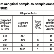
VERIS Molecular Diagnostic System sample-to-sample crossover contamination study
Summary
Sample carry-over and cross-contamination present a high risk for the laboratory. VERIS was developed to have a false positive rate due to cross contamination of less than 1 in 500 tests with an overall design goal of zero. This study involved several stages: to assess the sample-to-sample contamination rate using a real-time PCR assay and then characterize potential sources of contamination. System modifications were then to be developed to resolve any carry-over and cross-contamination and the system then retested.
Method and Results
Contamination characterization was performed by swabbing areas of the instrument before and after running a series of high concentration level positive samples to determine potential sources of contamination. Twenty high positive Cytomegalovirus (CMV) spiked samples at a concentration of 1×10¹⁰ IU/mL were used in this testing. A total of 27 high risk areas on the instrument were evaluated. High risk areas were defined as areas on the system where liquid handling occurs and where potential splashing of sample or reagents can occur. The swabs were placed in the TE buffer and then processed on the VERIS system. Swab assessments identified several areas where splashing and contamination of high positive samples was occurring, with the potential to contaminate a future sample.**
This resulted in several modifications to the liquid handling parameters and motor speeds to eliminate the potential for sample-to-sample contamination. Swab testing was used to verify the effectiveness of the modifications. The study concluded that accurate results for true negative samples were now being shown, with no detectable carryover and contamination from high positive to negative samples. This was true when the concentration of CMV target in the samples was above clinical levels and the frequency of high positives in the sample population exceeded 30%.
Cytomegalovirus (CMV) viral load assay for the VERIS MDx System
Summary
The initial assay menu includes the VERIS Cytomegalovirus (CMV) Assay*, intended for use in conjunction with clinical presentation and other laboratory markers as an aid in monitoring CMV viral load and for the detection of virus reactivation. This study reported on performance in key analytical and clinical measures.
Method and Results
137 paired samples tested on both the VERIS CMV Assay and the Roche COBAS AmpliPrep/COBAS TaqMan CMV Test were used to demonstrate method comparison in accordance with CLSI EP9-A2. 287 specimens were tested to demonstrate the clinical specificity of the assay.
Assay measuring interval: the assay is linear for human CMV with a lower limit of quantitation (LLQ) of 120 IU/mL and an upper limit of quantitation (ULQ) of 10,000,000 IU/mL. A nine member panel of the CMV AD169 reference strain demonstrated a linear range of 159 to 13,400,000 IU/mL (2.20-7.13 Log IU/mL). A four-member panel of the 1st WHO International Standard for Human Cytomegalovirus (HCMV) (NIBSC 09/162) demonstrated a linear range from 120 and 10,000 IU/mL (2.08-4.00 Log IU/mL)
Precision: demonstrated a total (within-run and between-run) standard deviation of less than 0.15 Log IU/mL across its linear range.
Sensitivity: The VERIS CMV Assay was shown to have a Limit of Detection (LoD) of 30 IU/mL (1.48 Log IU/mL) across all subtypes tested.
Performance evaluation of the Beckman Coulter VERIS Cytomegalovirus Assay on the VERIS MDx System
Summary
Another study assessed the VERIS CMV assay for reproducibility and specificity, comparing it with the Roche COBAS AmpliPrep/COBAS TaqMan CMV test. In immune-compromised individuals, the activation of the latent virus, is a significant cause of morbidity and mortality. Real-time polymerase chain reaction (PCR) assays offer the ability to diagnose active infection and monitor those individuals at risk.
Method and Results
Using paired plasma samples, total assay imprecision was
≤ 4.6% CV with the SD ≤0.14. Clinical specificity with negative samples was 100% with a lower bound of the 95% CI = 98.7%. It had comparable recoveries to the Roche assay with a Passing-Bablock regression equation of VERIS=0.30+1.00 (Roche), r=0.88 and n=130.
*Not for sale or distribution in the U.S.; not available in all markets.
**References available on request
November 2024
The leading international magazine for Clinical laboratory Equipment for everyone in the Vitro diagnostics
Beukenlaan 137
5616 VD Eindhoven
The Netherlands
+31 85064 55 82
info@clinlabint.com
PanGlobal Media is not responsible for any error or omission that might occur in the electronic display of product or company data.
This site uses cookies. By continuing to browse the site, you are agreeing to our use of cookies.
Accept settingsHide notification onlyCookie settingsWe may ask you to place cookies on your device. We use cookies to let us know when you visit our websites, how you interact with us, to enrich your user experience and to customise your relationship with our website.
Click on the different sections for more information. You can also change some of your preferences. Please note that blocking some types of cookies may affect your experience on our websites and the services we can provide.
These cookies are strictly necessary to provide you with services available through our website and to use some of its features.
Because these cookies are strictly necessary to provide the website, refusing them will affect the functioning of our site. You can always block or delete cookies by changing your browser settings and block all cookies on this website forcibly. But this will always ask you to accept/refuse cookies when you visit our site again.
We fully respect if you want to refuse cookies, but to avoid asking you each time again to kindly allow us to store a cookie for that purpose. You are always free to unsubscribe or other cookies to get a better experience. If you refuse cookies, we will delete all cookies set in our domain.
We provide you with a list of cookies stored on your computer in our domain, so that you can check what we have stored. For security reasons, we cannot display or modify cookies from other domains. You can check these in your browser's security settings.
.These cookies collect information that is used in aggregate form to help us understand how our website is used or how effective our marketing campaigns are, or to help us customise our website and application for you to improve your experience.
If you do not want us to track your visit to our site, you can disable this in your browser here:
.
We also use various external services such as Google Webfonts, Google Maps and external video providers. Since these providers may collect personal data such as your IP address, you can block them here. Please note that this may significantly reduce the functionality and appearance of our site. Changes will only be effective once you reload the page
Google Webfont Settings:
Google Maps Settings:
Google reCaptcha settings:
Vimeo and Youtube videos embedding:
.U kunt meer lezen over onze cookies en privacy-instellingen op onze Privacybeleid-pagina.
Privacy policy