Beukenlaan 137
5616 VD Eindhoven
The Netherlands
+31 85064 55 82
info@clinlabint.com
PanGlobal Media is not responsible for any error or omission that might occur in the electronic display of product or company data.
Sepsis is a life threatening inflammatory disorder and the immune systems response to infection. It is one of the leading causes of death in hospitalized patients worldwide with 1.8 million cases annually. Improvement in survival remains contingent on early recognition of the causative organism to enable targeted antimicrobial therapy.
by Kelly Marie Ward and Rhian Harris
Sepsis incidence
Sepsis is a life threatening inflammatory disorder and the immune systems response to infection [1]. It is one of the leading causes of death in hospitalized patients worldwide with 1.8 million cases annually [2]. Each year, 37 000 deaths are caused by sepsis in the UK [2–4]. Mortality rates remain between 25–30% for severe sepsis and 40–70% for septic shock, despite advances in pharmacotherapy and supportive care [1] and various campaigns, e.g. the Surviving Sepsis Campaign (SSC) [3]. This is mainly due to poor identification and delayed interventions [3]. Data from the SSC showed a mortality rate of 39.8% among 15 022 patients and 39.8% of those admitted to critical care in England and Wales die in hospital [2]. A hospital admission with severe sepsis places the patient at a level of risk 6–10-fold greater than admission with an acute myocardial infarction and 4–5 times greater than if they had suffered an acute stroke [2].
What is sepsis?
The American College of Chest Physicians and the Society of Critical Care Medicine classified the continuum of an inflammatory response to microorganisms as ‘systemic inflammatory response syndrome’ (SIRS) [1]. SIRS is a collection of signs that show the body is reacting to a range of injuries or illnesses and it is not specific to infection [3]. It is identified when two of the following symptoms – fever, tachycardia, tachypnea and leukopenia are met in the absence of an infection [3].
Uncomplicated sepsis is the presence of an infection in association with SIRS [1] in the absence of organ dysfunction [4]. Bacteria that cause infection can enter the body via breaks in the skin, catheters and underlying infections in the urinary, respiratory or gastrointestinal tract [5]. Sepsis can be defined as ‘a systemic disease that is caused by the spread of microorganisms and their toxins via the circulating blood’ [6]. The endo and exotoxins produced by different organisms often lead to an inflammatory response of varying severities [7]. Severe sepsis occurs when SIRS is accompanied by infection and organ dysfunction [4]. Figure 1 taken from Royal College of Physicians: Acute Care Toolkit 9: Sepsis; September 2014 [4] demonstrates this balance.
Pathophysiology of sepsis
The pathophysiology of sepsis involves a complex interaction of proinflammatory and anti-inflammatory mediators in response to pathogen invasion [1]. When an infectious agent invades the host, an innate response is triggered via toll-like receptors (TLR) [8]. These are trans-membrane proteins with the ability to promote signalling pathways downstream and trigger cytokine release, neutrophil activation and stimulation of endothelial cells [8]. The cytokines such as interleukin (IL)-1 and IL-6 are released from the cells where inflammatory reactions have commenced. They stimulate lymphocytes and mononuclear cells to produce further cytokines, resulting in the recruitment and migration of further cells to the site or organ where inflammation is occurring [9]. This leads to endothelium damage, vascular permeability, microvascular dysfunction, coagulation pathway activation and impaired tissue oxygenation resulting in the cascade of sepsis [1]. There is activation of humoural and cell-mediated immunity with specific B and T cell responses and both pro and anti-inflammatory cytokine release [8]. Adaptive immunity is triggered and the inflammatory cascade of sepsis occurs where the balance is shifted towards cell death and a state of relative immunosuppression and end organ dysfunction ensues with hemodynamic changes causing elevated cardiac output and generalized vasodilation described as shock [8]. As the inflammatory response progresses, myocardial depression is more pronounced resulting in a falling cardiac output. There is capillary leak and pulmonary edema that may progress to acute lung injury. Renal failure then follows accompanied by alterations in the coagulation cascade towards a pro-coagulant and antifibrinolytic state. The development of ‘disseminated intravascular coagulation’ (DIC) in severe sepsis is a predictor of death and the development of multiorgan failure [8]. The respiratory, genitourinary and gastrointestinal systems are most commonly infected and pneumonia is the most common presentation leading to sepsis [1].
Clinical presentation and diagnosis
There are a variety of symptoms that can indicate sepsis, including fever, chills, decreased blood pressure, shaking, skin rash, confusion and a rapid heartbeat [10].The clinical diagnosis of sepsis is most often made before culture results are available and although localized signs and symptoms may be present, organ hypoperfusion or shock can occur without the knowledge of the cause [1]. Fever is the most common manifestation of sepsis and 40% of those patients will have hypotension [1].
A vast array of laboratory tests are required for the diagnosis and management of sepsis including full blood counts, basic metabolic panels, lactate and liver enzyme levels and C-reactive protein. In the Microbiology laboratory we would expect to receive, blood cultures: two peripheral and from each indwelling catheter, urine, stools if symptoms of diarrhoea, sputum and skin and soft tissue for culture if clinically significant [1] Currently blood cultures are the definitive diagnosis tool when septicemia is suspected [11]. Blood culture systems have evolved over time to ensure optimum isolation of any organisms present by adding different nutrients, introduction of automated systems and increasing the detection rate of a positive as a result of new software [12]. They are used to detect the presence of any microorganisms present by providing optimum conditions for growth. However in 50–65% of patients the blood culture is often negative [1].
Early management and identification of infectious cause
Due to the high mortality rates the early identification and management of sepsis is crucial and requires respiratory stabilization followed by fluid resuscitation, vasopressor therapy, infection identification and control and prompt antibiotic administration [1].
The SSC published ‘The Resuscitation Bundle’ which comprised a set of tasks to be to be completed within the first 6 hours after the clinical identification of sepsis [2]. The first four tasks were:
These tasks involve the Pathology laboratory and it was established that systems within healthcare environments needed to be well designed and implemented to ensure that the appropriate investigations, equipment and treatments were available at the point of care [2].
The length of time that it takes for the correct identification of the causative organism has many ramifications both clinically and financially. Empiric antibiotic therapy is based on the most likely source, clinical context, recent antibiotic use and local resistance patterns. This should be narrowed when the causative agent has been identified to reduce the risk of resistance or superinfection [1]. The length of time the patient is prescribed such antibiotics may be reduced if the causative organism is characterized sooner, and inappropriate therapy changed accordingly. The use of targeted narrow spectrum antibiotics might reduce bed days resulting in a financial saving. Using antibiotics more than is required in both humans and animals has resulted in the increased emergence of antibiotic resistance [13]. New antibiotics are produced very slowly and as a result it is important to limit any likelihood of the spread of resistance between organisms by only prescribing the antibiotic necessary [14]. Also early appropriate antibiotic therapy is associated with improved clinical outcomes [1]. The antibiotics should be administered within 1 hour of suspected sepsis. In septic shock early antibiotic therapy increases survival and for each hour this is delayed the survival rates decrease by 8% [1]. The possible benefits to the patient in terms of improved treatment of infections, the potential to reduce costs and an attempt to reduce the emergence of antibiotic resistance have led to the development and research into more rapid diagnosis techniques.
MALDI-TOF and the Bruker Sepsityper blood culture kit
Matrix-assisted laser desorption/ionization–time of flight (MALDI-TOF) spectroscopy can identify organisms from intact cells based on the profile of different proteins and relative molecular mass [15]. A smear of the cultured organism is placed onto a stainless steel target plate, with matrix placed over the top. Matrix is used as it prevents fragmentation of higher mass molecules. The laser is fired at the smear generating a cloud of ions which are accelerated up the flight tube to the detector, where the time of flight is converted to Daltons (Da)/molecular mass. The heavier the molecular mass of the ion the greater the time of flight (Fig. 2).
This is also known as proteomic profiling. A spectrum is produced for each organism based on their mass/charge ratio, which is determined by the different molecular mass and charge of the ions present for the organism in question [15]. A spectrum is produced with a variety of peaks each one representing a different molecular fragment which has been released as a result of the laser desorption [15]. This spectrum is then compared to the database for possible matches and is scored based on the number of peaks that match the corresponding organism (Fig. 3).
This method can be used to identify bacteria, yeasts, moulds, mycobacteria and Nocardia to species level using species-specific spectral patterns [16]. Using this method, identification from a bacterial culture can be achieved in 30–60 seconds.
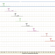
MALDI-TOF spectroscopy can also be used to identify organisms directly from blood culture bottles using the Sepsityper kit extraction method (Fig. 4). This takes approximately 30 minutes and allows accurate identification to species level on day 1 of the bottle being flagged as positive. The use of the Sepsityper kit could enhance task three of the SSC Resuscitation bundle by allowing earlier targeted therapy. Components within the blood culture such as red cells, white blood cells and serum can interfere with the analysis resulting in the formation of additional spectral peaks [17]. These peaks will not be found in the database and will result in difficulty in interpreting the results, which is why the extraction kit by Bruker has been developed. The development of this kit has allowed purification and extraction to be carried out to optimize recovery of the bacteria present in the blood culture sooner. This is carried out by a series of centrifugation steps to separate any organisms present from the blood and fluid present in the blood culture and also formic acid to breakdown the cell wall of the organism to aid identification using MALDI-TOF technology (Bruker – Introduction for use Maldi Sepsityper kit (Accessed 2015).
Our laboratory evaluated the use of the Sepsityper kit to identify the causative organism direct from the positive blood culture bottle using MALDI-TOF spectroscopy. The results were retrospectively analysed to determine if there would have been a change to the antibiotic therapy if this method was in routine use.
Study results
Two hundred and thirty-six positive blood cultures were analysed retrospectively and compared against current laboratory methods. The results are shown in Table 1.
Table 1 shows the percentage of successful identifications achieved by using the Bruker Sepsityper method. The percentage of blood cultures that achieved successful identification within 1 hour of becoming positive was 75.42% (green plus yellow rows). A score of above 1.8 indicates a secure genus and probable species identification (green row) [18], a score between 1.6 and 1.8 indicates probable species identification (yellow row) [18]. Any score below 1.6 cannot be accepted as a reliable identification (red row). There was a 93.33% agreement of identification between the Bruker Sepsityper kit or direct MALDI-TOF identification versus the BD Phoenix and other conventional laboratory methods.
The previous antibiotic treatment, the clinical history of the patient and the identification of the organism produced by the Bruker Sepsityper kit on day one was analysed retrospectively by the consultant microbiologist to determine if there would have been any clinical impact if the identification of the organism had been known on day 1. As the organism that is causing the infection is not known immediately, patients are started on broad-spectrum or a combination of antibiotics when bacteremia is suspected; however, incorrect or insufficient therapy has been associated with increased mortality, morbidity and increased hospital stay [19]. The consultant microbiologist determined that 26 (11%) out of the 236 blood cultures analysed would have indicated a requirement for the patient to have their antibiotic therapy altered in some way. Sixteen of the 26 positive blood cultures indicated that the patients’ antibiotic therapy could be reduced from a broad-spectrum antibiotic to a narrower spectrum antibiotic. This can have huge cost savings implications as well as reduce the likelihood of resistance emerging against broad-spectrum antibiotics [20]. Knowing the identification of the organism on the day the blood culture bottle is flagged as positive enables the antimicrobial therapy to be changed accordingly therefore helping to reduce the emergence of resistance, provide targeted therapy for better treatment outcomes and reduce bed days spent in hospital. Improvement in survival remains contingent on the early recognition and management of severe sepsis and septic shock [1].
References
1. Gauer RL. Early Recognition and Management of Sepsis in Adults: The First Six Hours. Am Fam Physician. 2013; 88: 44–53.
2. Daniels R. Surviving the First Hours In Sepsis: getting the basics right (an intensivists perspective). J Antimicrob Chemother. 2011; 66(Suppl 2): ii11–23.
3. McClelland H, Moxon A. Early identification and treatment of sepsis. Nursing Times 2014; 110: 14–17. (http://www.nursingtimes.net/Journals/2014/01/17/q/v/z/220114-Early-identification-and-treatment-of-sepsis.pdf)
4. Royal College of Physicians. Acute Care Toolkit 9: Sepsis; September 2014. (https://www.rcplondon.ac.uk/sites/default/files/acute_care_toolkit_9_sepsis.pdf)
5. Public Health England. Investigation of blood cultures. Bacteriology 2014; B37(8). (https://www.gov.uk/government/uploads/system/uploads/attachment_data/file/372070/B_37i8.pdf)
6. Odeh M. Sepsis, septicaemia, sepsis syndrome, and septic shock: the correct definition and use. Postgrad Med J. 1996; 72(844): 66.
7. Martin GS. Sepsis, severe sepsis and septic shock: changes in incidence, pathogens and outcomes. Expert Rev Anti-infect Ther. 2012; 10(6): 701–706.
8. Ventetuolo CE, Levy MM. Sepsis: a clinical update. Clin J Am Soc Nephrol. 2008; 3: 571–577.
9. Roitt I, Brostoff J, Male D. Cell migration and inflammation. In: Cook L, Immunology, 4th ed. Mosby 1998.
10. Severe sepsis/septic shock, recognition and treatment protocols. Stony Brock Medicine 2013. (http://www.survivingsepsis.org/sitecollectiondocuments/protocols-sepsis-treatment-stony-brook.pdf)
11. Previsdomini M, Gini M, et al. Predictors of positive blood cultures in critically ill patients: a retrospective evaluation. Croat Med J. 2012; 53(1): 30–39.
12. Zadroga R, Williams DN, et al. Comparison of 2 blood culture media shows significant differences in bacterial recovery for patients on antimicrobial therapy. Clin Infect Dis. 2012; 56(6): 790–797.
13. Rao GG. Risk factors for the spread of antibiotic-resistant bacteria. Drugs 1998; 55(3): 323–330.
14. Guidos RJ. Combating antimicrobial resistance: policy recommendations to save lives. Clin Infect Dis. 2011; 52(5): 397–428.
15. Carbonnelle E, Beretti JL, et al. (2007). Rapid identification of Staphylococci isolated in clinical microbiology laboratories by matrix-assisted laser desorption ionization-time of flight mass spectrometry. J Clin Microbiol. 2007; 45(7): 2156–2161.
16. Stevenson LG, Drake SK, Murray PR. Rapid identification of bacteria in positive blood culture broths by matrix-assisted laser desorption ionization-time of flight mass spectrometry. J Clin Microbiol. 2010; 48(2): 444–447.
17. Lagacé-Wiens PRS, Adam HJ, et al. Identification of blood culture isolates directly from positive blood cultures by use of matrix-assisted laser desorption ionization–time of flight mass spectrometry and a commercial extraction system. J Clin Microbiol. 2012; 50(10), 3324–3328.
18. El-Bouri K, Johnston S, et al. Comparison of bacterial identification by MALDI-TOF mass spectrometry and conventional diagnostic microbiology methods: agreement, speed and cost implications. Br J Biomed Sci 2012; 69(2): 47–55.
19. Kollef MH. Broad-spectrum antimicrobials and the treatment of serious bacterial infections: getting it right up front. Clin Infect Dis. 2008; 47(1): 3–13.
20. Rüttimann S, Keck B, et al. Long-term antibiotic cost savings from a comprehensive intervention program in a medical department of a university-affiliated teaching hospital. Clin Infect Dis. 2004; 38(3): 348–356.
The authors
Kelly Marie Ward* MSc, FIBMS; Rhian Harris MSc, AIBMS
Royal Glamorgan Hospital Microbiology Laboratory, Cwm Taf University Health Board, Llantrisant, Glamorgan, UK
*Corresponding author
E-mail: Kelly.Ward@wales.nhs.uk
Since the mapping of the human genome was completed over a decade ago, our knowledge of genetic drivers of disease continues to evolve at an ever-quickening pace. Consequently, genetic testing and pharmacogenomics have become common within the healthcare system and have generated the knowledge that has empowered us to both understand and influence our lifelong health through pre-emptive intervention.
Progress in medical genomics and its impact on healthcare cannot be understated; from genotyping patients to predict drug response, to stratifying patients according to the risk of a disease, molecular testing is having a very positive impact on many patient treatment pathways.
Undoubtedly, we are now more aware and in control of our health than ever before. It is no surprise then that the molecular diagnostic market has become the fastest growing segment of the IVD industry with assays serving the gamut of disease areas and breaking new boundaries in personalized healthcare. Despite the public appetite and availability of powerful molecular diagnostic assays that can unequivocally diagnose genetic disorders, their use has not gained universal acceptance. Many traditional diagnostic tests continue to under-diagnose, or diagnostic testing is not attempted, leading to missed opportunities for early and appropriate therapy intervention of potentially life-threatening diseases. One prime example where a molecular diagnostic approach can improve health is mutation profiling for Familial Hypercholesterolemia (FH).
Familial Hypercholesterolemia
Familial Hypercholesterolemia (FH) is a genetic disorder of lipoprotein metabolism. It is a common autosomal dominant, or inherited, disease which affects the plasma clearance of LDL-cholesterol (LDL-C), resulting in premature onset of cardiovascular disease (CVD) and a higher mortality risk.
Early diagnosis of FH is very advantageous as by the time heterozygous FH sufferers enter early adulthood they will have accumulated years of continuous build-up of fatty or lipid masses in arterial walls and are at one hundred-fold greater risk of a heart attack than their non-FH peers. If left untreated, men and women with heterozygous FH with total cholesterol levels of 8–15 mmol/L typically develop coronary heart disease (CHD) before age 55 and 60, while homozygotes with total cholesterol levels of 12–30 mmol/L typically develop CHD very early in life and if untreated die before age 20.
Clinical diagnosis of FH relies on five criteria: family history, clinical history of premature CHD, physical examination for xanthomas and corneal arcus, very high LDL cholesterol on repeated measurements, and / or a causative mutation detected by molecular genetics. To formally quantify this, a number of sets of statistically and genetically validated criteria have been devised; namely the Dutch Lipid Clinic Network Criteria and the Simon Broome Criteria. These classify suspected cases into definite, possible and probable diagnoses of FH. In the absence of definitive diagnosis through detection of a causative mutation using molecular genetics, clinical diagnosis could miss a considerable proportion of FH patients, particularly those with a mild phenotype and the pediatric population in whom the phenotype has not appeared yet.
The UK, US and international guidelines now recommend that probable or possible FH patients undergo a DNA test to confirm the diagnosis of FH. Recommendations also advocate that once an activating mutation has been found in one family member (the index case), cascade screening of that mutation in first degree relatives of the index case should proceed. Cascade screening using a molecular assay can thus identify index family members who may otherwise be asymptomatic.
The good news is that if detected early, FH can be treated successfully with lipid lowering therapy and lifestyle changes. In comparison to other hyperlipidemias, FH therapy tends to be more aggressive, so definitive diagnosis has additional benefits in determining care packages. Statin drug therapy significantly reduces the morbidity and mortality from premature coronary disease in FH, particularly if affected individuals are identified and treated in childhood or early adulthood. Accurate and early diagnosis of specific mutations can result in a better overall outcome for patients through the prescribing of tailored treatments to reduce morbidity and mortality from premature cardiovascular disease. Different mutations can dictate different directions of management, such as the poorer response to lipid-lowering therapy with certain LDLR mutations. The identity of the gene involved can potentially aid the clinician to decide on how aggressive the treatment strategy will be.
Mutation diagnosis also provides clarity, and can help with an individual’s understanding and acceptance of their condition. Also a greater compliance with cholesterol lowering medication is observed with those who have been genetically diagnosed with FH.
Mutational profiling of FH
Currently, ~1200 mutations have been documented worldwide in LDLR; these affect all functional domains of the LDL receptor protein and include single-nucleotide mutations, copy number variations, and splicing mutations throughout the LDLR gene. A single mutation, Arg3500Gln, is the only common FH-related mutation in APOB, while c.1120G>T mutation is predominately detected in PCSK9. Heterozygous LDLR, APOB, and PCSK9 mutations are found in >90%, ~5%, and ~1%, respectively, of heterozygous FH subjects with a causative mutation. Prevalence varies geographically.
The abundance of different FH mutations can make genetic testing labour-intensive and costly, with many laboratories defaulting to performing expensive and lengthy Next Generation Sequencing (NGS) tests in an effort to ensure a comprehensive mutational screen. However, as our understanding of the genetic drivers of FH, as well as common population-specific mutations, increases, novel assays and techniques are being developed to meet the needs facing clinical genetics laboratories, including cost, throughput and time to result.
Randox Laboratories have developed The Familial Hypercholesterolaemia (FH) Arrays I and II that are rapid, simple and accurate diagnostic tests which enable simultaneous detection of 40 FH-causing mutations (20 mutations per array) within the LDLR, ApoB and PCSK9 genes. The assay is based on multiplex PCR followed by biochip array hybridization. Using mutation rate data from a study of 500 UK and Ireland families with genetically-confirmed FH, the Randox FH Arrays are capable of detecting approximately 71% of activating mutations in this population. The mutations will also be detected in other geographical regions.
The assay can be completed from extracted DNA to an easy-to-interpret result report in 3 hours, with the requirement for only 20ng of genomic DNA per array. The system can be used to detect small base changes, insertions and deletions within the same multiplex PCR, allowing addition of new FH mutational targets if required. The arrays are designed for use on the Evidence Investigator (Randox Laboratories Limited, Crumlin, UK). This instrument has been developed alongside Randox’s proprietary Biochip Array Technology (BAT), a multiplex testing platform founded on ELISA principles that currently has application within clinical immunoassays, drug development R&D, clinical research, forensic and clinical toxicology, veterinary drug residues and molecular diagnostics.
FH Array I and II workflow
Randox’s multiplex assays, such as FH Array I and II, have been specifically designed to detect the most common mutations, provide a cost-effective and clinically relevant alternative to NGS testing. Targeting the most commonly detected mutations in a given population enables diagnosis within hours rather than months. Where a mutation is identified in an index patient, cascade testing of family members only requires the mutation in question to be targeted; therefore negating the use of broad profiling approaches such as NGS in this setting.
Conclusion
FH is a common yet underdiagnosed condition that poses a significant risk to public health worldwide. In 2008, cardiovascular diseases were the leading cause of non-communicable deaths worldwide, with an estimated mortality rate of 17 million people. Raised cholesterol was attributed to 2.6 million deaths. Understanding a person’s genetic predisposition to cardiovascular disease through genetic testing will allow patients to receive appropriate therapeutic and interventional treatment to reduce morbidity and mortality associated with cardiovascular disease.
Pioneering multiplex diagnostic assays, tailored to incorporate the relevant FH-causing mutations, provide a promising future for both genetic laboratories, where a rapid, cost-effective approach to determine mutational status in cases of suspected FH is enabled, and the patient, whose treatment and care pathway is managed effectively.
The author
Martin Crockard, PhD
Randox Laboratories Ltd.
55 Diamond Road, Crumlin, Co. Antrim
U.K.
Early diagnosis of sepsis is essential for enabling appropriate treatment. PCT and MR-pro ADM have been shown to be independent biomarkers for sepsis and progression to septic shock, and simultaneous analysis seems to be more effective than the single marker approach.
by Dr S. Angeletti, M. De Cesaris, Dr A. Lo Presti, et al.
Introduction
Sepsis is a severe condition that represents the tenth most common cause of death in the USA. In Europe, sepsis occurs in more than 35% of the patients admitted in the intensive care unit. The mortality associated with sepsis is approximately 28% and it rises to 40–60% in cases of septic shock, despite adequate treatment administration. Nearly 9% of patients with sepsis experience severe sepsis and nearly 3% progress to septic shock leading to multi-organ failure. More than 50% of patients affected by septic shock do not survive [1–3]. Consequently, the rapid recognition and treatment of sepsis is mandatory to reduce both the mortality and the hospitalization with related costs [1-3].
Sepsis is commonly defined as the presence of infection in conjunction with the systemic inflammatory response syndrome (SIRS); severe sepsis, as sepsis complicated by organ dysfunction; and septic shock, as sepsis-induced acute circulatory failure characterized by persistent arterial hypotension despite adequate volume resuscitation and not explained by other causes [1, 4]. The diagnosis of sepsis and evaluation of its severity is complicated by the highly variable and non-specific nature of the signs and symptoms of sepsis [5]. However, the early diagnosis and stratification of the severity of sepsis is very important, increasing the possibility of starting timely and specific treatment [4, 6].
The gold standard for detection of bloodstream infections is blood culture. The time required for a positive blood culture result depends on the incubation time required for the culture to turn positive and the subsequent biochemical identification, along with an antibiotic sensitivity test, both of which usually take 48 h [7]. Furthermore, in some cases, blood culture results remain negative owing to empirical broad-spectrum antibiotics that are frequently started in the presence of SIRS and often continued for a prolonged time course despite the absence of clinical and microbiological data supporting a diagnosis of bacterial infection [4, 8]. Several studies have evaluated the diagnostic utility of various biomarkers, including ferritin, haptoglobin, interleukin 6, C-reactive protein (CRP) and procalcitonin (PCT) for suspected sepsis in the ICU patient population [9–11].
It remains difficult to differentiate sepsis from other non-infectious causes of SIRS [12] and there is a continuous search for better biomarkers of sepsis.
PCT is a polypeptide that has demonstrated the highest reliability in the early diagnosis of sepsis, severe sepsis or septic shock compared to other plasma biomarkers or clinical data alone [13]. Moreover, PCT has been advocated also to clarify the bacterial origin of some localized infections [14–15].
The mid-regional pro-adrenomedullin (MR-proADM) has been shown to play a decisive role in both the induction of hyper-dynamic circulation during the early stages of sepsis and the progression to septic shock [16–18], and recently it has been reported that MR-proADM differentiates sepsis from non-infectious SIRS with high specificity. Moreover, simultaneous evaluation of MR-proADM and PCT in septic patients increased the post-test diagnostic probabilities compared to the independent determination of individual markers [19–20]; probably the multimarker approach seems to be the more effective [14, 19].
The aim of the present study was to perform a focused evaluation of the role of the combination of PCT and MR-proADM in patients with severe sepsis and septic shock (SS) to differentiate it from patients with mild sepsis or SIRS for a prompt and specific treatment administration.
Methods
Patient and control characteristics
One hundred and seventeen patients with SS and 100 patients with SIRS, hospitalized at the University Hospital Campus Bio-Medico of Rome between the years 2012 and 2014, were enrolled in the study. The patients’ details are reported in Table 1.
Sepsis was defined by the American College of Chest Physicians/Society of Critical Care Medicine Consensus Conference definition of sepsis [4] based on the presence of a recognized site of infection and evidence of a SIRS occurring when at least two of the following criteria are present: body temperature higher than 38°C or lower than 36°C, heart rate higher than 90 beats per minute, respiratory rate higher than 20 breaths per minute or hyperventilation as indicated by an arterial partial pressure of carbon dioxide (PaCO2) lower than 32 mm Hg and a white blood cell count of higher than 12,000 cells/mm3 or lower than 4,000.
Patients were classified according to clinical signs into SS and SIRS. Acute physiological and chronic health evaluation (APACHE) II and sequential organ failure assessment (SOFA) scores were computed. APACHE II scores in SS and SIRS patients were calculated by Medscape, APACHE II scoring system calculator [21]. The SOFA score was calculated only for SS patients to better define the severity of the sepsis [22–23]. The study was approved by the Ethics Committee of the University Hospital Campus Bio- Medico, Rome, Italy.
Blood culture
Blood samples for blood culture were collected when patients showed the symptoms and signs of SIRS [1, 2, 4]. Blood culture included three sets (time 0, time 30 and time 60 min) of one aerobic and one anaerobic broth bottles (Bactec Plus Aerobic/F, Bactec Plus Anaerobic/F, Beckton Dickinson) per patient drawn during 1-h period of clinically suspected bloodstream infection. Blood culture vials were incubated in the Bactec 9240 automated system (Beckton Dickinson). Blood culture samples that turned positive were immediately processed for Gram staining and cultivated. Bacterial identification was performed by MALDI-TOF, as previously described [24].
PCT and MR-proADM measurement
The plasma concentrations of PCT and MR-proADM were measured by an automated analyser using a time-resolved amplified emission method (Kryptor, Brahms AG), with commercially available assays (Brahms AG) [25].
Statistical analysis
Data was analysed using MedCalc 11.6.1.0 statistical package (MedCalc Software). Plasma levels of PCT and MR-proADM were log-transformed to achieve a normal distribution. The normal distribution of each marker concentration was tested by the Kolmogorov–Smirnov test. PCT and MR-proADM in patients with SIRS and SS were compared using the Mann–Whitney test. Multiple logistic regression analysis (stepwise method) using SS versus PCT and MR-proADM was performed and the odds ratio (OR) computed. For OR calculation variables were retained for P<0.05 and removed for P>0.1.
Receiver operating characteristic (ROC) analysis was performed among independent variables associated with SS to define the cut-off point for plasma PCT and MR-proADM and their diagnostic accuracy to predict SS [26]. Pre-test odds, post-test odds and the consequent post-test probability were computed to investigate whether the combination of PCT and MR-proADM improves post-test probability. Likelihood ratios were used as these tests are not prone to bias due to prevalence rates [27].
Results
Patients with SS and SIRS characteristics
The mean age of the 117 patients with SS (71 men and 46 women) was 69 ± 3 years (Table 1). The principal comorbidities of patients with SS and SIRS and the sources of bacteremia are summarized in Table 1. In patients with SS the average APACHE II score value was 19.8, corresponding to 24% risk of death and the average SOFA score was 6.8 corresponding to a predicted mortality of <33%. In patients with SIRS the APACHE II score was 7, corresponding to 6% risk of death (Table 1).
SS was caused by Gram-negative pathogens in 63/117 (54%) of patients and in Gram-negative sepsis, E. coli (28/63; 44.4%) was the most frequent isolate. Gram-positive SS was present in 24/117 (20.5%) of cases and the most frequent pathogen was S. aureus (14/24; 58.3%), whereas C. albicans was the most frequent isolate in yeast-positive cultures (10/117; 8.5%) and blood cultures were polymicrobial in 20/117 (17%) cases. Bacterial isolates from positive blood culture are reported in Table 2.
PCT and MR-proADM in patients with SS and SIRS
Median values, interquartile ranges (25th percentile and 75th percentile) and Mann–Whitney comparison of PCT and MR-proADM analysed in patients with SS and SIRS are reported in Table 3. PCT and MR-proADM values were significantly higher in patients with SS than SIRS (P<0.0001) (Table 3 and Figure 1).
ROC curve and AUC analysis of PCT and MR-proADM in patients with SS
In SS patients, the area under curve (AUC) values of PCT and MR-proADM are reported in Table 4. Based upon ROC curve analysis and AUC characteristics, PCT and MR-proADM were considered applicable for sepsis diagnosis at the cut-off values of 0.5 ng/mL and 1 nmol/L, respectively (Table 4 and Figure 2).
Multiple logistic regression analysis
Multiple logistic regression analysis using SS as the dependent variable and PCT and MR-proADM as independent variables is reported in Table 5. Patients with MR-proADM >1 nmol/L have ~195 times the probability of being affected by SS than patients with SIRS, and patients with PCT values >0.5 ng/mL have the probability of developing SS 49 times more than SIRS.
Combined PCT and MR-proADM measurement in SS diagnosis: post-test probability calculation
In patients with SS, PCT and MR-proADM used as single markers have a post-test probability of 0.964 and 0.936, respectively. The combination of PCT and MR-proADM resulted in a higher value of post-test probability, 0.996 (Table 4).
Discussion
The early diagnosis and stratification of the severity of sepsis are essential, increasing the possibility of starting timely the specific treatment, especially in patients affected by SS. In this study, the combined measurement of PCT and MR-proADM in patients with SS was evaluated in order to establish the advantage derived from the use of a multimarker rather than a single marker approach.
PCT has been described as a reliable marker in the early diagnosis of sepsis compared to other plasma biomarkers or clinical data alone [13, 14, 19]. MR-proADM has been used as marker of disease severity in different clinical setting and recently its combination with PCT in bacterial infections and sepsis has been evaluated [28–32, 14, 19]. The combination of PCT and MR-proADM could allow the simultaneous evaluation of the presence of a bacterial infection as well as of the severity of this infection, giving to the ward clinicians a first useful indication waiting for blood culture positivity.
Results from this study demonstrated that in patients with SS, PCT and MR-proADM values are significantly higher than patients with SIRS. ROC curve analysis of PCT and MR-proADM demonstrated a high diagnostic accuracy of these two markers in SS diagnosis at the cut-off value of 0.5 ng/mL and 1 nmol/L, respectively. The logistic regression analysis showed higher OR values for both markers indicating a significant increased risk of having SS when these markers are higher than the cut-off values established. Furthermore, the combination of the two markers leads to a very high post-test probability value of about 99.6%.
These data confirmed the important role of the combination of PCT and MR-proADM in the diagnosis and prognosis of patients with sepsis rather than the single marker approach, because it combines the diagnostic ability of PCT with the prognostic value of MR-proADM, as already described in localized bacterial infections and not complicated sepsis [14, 19].
In conclusion, this study further support the advantage derived from the multi-marker approach in sepsis diagnosis and prognosis, especially in critically ill patients.
References
1. Dellinger RP, Levy MM, Rhodes A, Annane D, Gerlach H, et al. Surviving Sepsis Campaign: international guidelines for management of severe sepsis and septic shock, 2012. Intensive Care Med. 2013; 39: 165–228.
2. Levy MM, Fink MP, Marshall JC, Abraham E, Angus D, et al. 2001 SCCM/ESICM/ACCP/ATS/SIS international sepsis definitions conference. Crit Care Med 2003; 31: 1250–1256.
3. Angus DC, Linde-Zwirble WT, Lidicker J, Clermont G, Carcillo J, Pinsky MR. Epidemiology of severe sepsis in the United States: analysis of incidence, outcome, and associated costs of care. Crit Care Med. 2001; 29: 1303–1310.
4. American College of Chest Physicians/Society of Critical Care Medicine. American College of Chest Physicians/Society of Critical Care Medicine consensus conference: definitions for sepsis and organ failure and guidelines for the use of innovative therapies in sepsis. Crit Care Med. 1992; 20: 864–874.
5. Martin GS, Mannino DM, Eaton S, Moss M. The epidemiology of sepsis in the United States from 1979 through 2000. N Engl J Med. 2003; 348: 1546–1554.
6. Russell JA. Management of sepsis. N Engl J Med. 2006; 355: 1699–1713.
7. Cherkaoui A, Hibbs J, Emonet S, Tangomo M, Girard M, et al. Comparison of two matrix-assisted laser desorption ionization time of flight mass spectrometry methods with conventional phenotypic identification for routine identification of bacteria to the species level. J Clin Microbiol. 2010; 48: 1169–1175.
8. Gullo A, Bianco N, Berlot G. Management of severe sepsis and septic shock: challenges and recommendations. Crit Care Clin. 2006; 22: 489–501.
9. Biomarkers Definitions Working Group. Biomarkers and surrogate endpoints: preferred definitions and conceptual framework. Clin Pharmacol Ther. 2001; 69: 89–95.
10. Marshall JC, Reinhart K. Biomarkers of sepsis. Crit Care Med. 2009, 37: 2290–2298.
11. Nakamura A, Wada H, Ikejiri M, Hatada T, Sakurai H, et al. Efficacy of procalcitonin in the early diagnosis of bacterial infections in a critical care unit. Shock 2009, 31: 591.
12. Vänskä M, Koivula I, Jantunen E, Hämäläinen S, Purhonen AK, et al. IL-10 combined with procalcitonin improves early prediction of complications of febrile neutropenia in hematological patients. Cytokine 2012; 60: 787–792.
13. Assicot M, Gendrel D, Carsin H, Raymond J, Guilbaud J, Bohuon C. High serum procalcitonin concentrations in patients with sepsis and infection. Lancet 1993; 341: 515–518.
14. Angeletti S, Spoto S, Fogolari M, Cortigiani M, Fioravanti M, et al. Diagnostic and prognostic role of procalcitonin (PCT) and MR-pro-Adrenomedullin (MR-proADM) in bacterial infections. APMIS 2015; 123: 740–748.
15. Kojic D, Siegler BH, Uhle F, Lichtenstern C1, Nawroth PP, et al. Are there new approaches for diagnosis, therapy guidance and outcome prediction of sepsis? World J Exp Med. 2015; 5: 50–63.
16. Hirata Y, Mitaka C, Sato K, Nagura T, Tsunoda Y, et al. Increased circulating adrenomedullin, a novel vasodilatory peptide, in sepsis. J Clin Endocrinol Metab. 1996; 81: 1449–1153.
17. Kitamura K, Kangawa K, Kawamoto M, Ichiki Y, Nakamura S, et al. Adrenomedullin: a novel hypotensive peptide isolated from human pheochromocytoma. Biochem Biophys Res Commun. 2012; 425: 548–555.
18. Zhou M, Ba ZF, Chaudry IH, Wang P. Adrenomedullin binding protein-1 modulates vascular responsiveness to adrenomedullin in late sepsis. Am J Physiol Regul Integr Comp Physiol. 2002; 283: R553–560.
19. Angeletti S, Battistoni F, Fioravanti M, Bernardini S, Dicuonzo G. Procalcitonin and mid-regional pro-adrenomedullin test combination in sepsis diagnosis. Clin Chem Lab Med. 2013; 51: 1059–1067.
20. Suberviola B, Castellanos-Ortega A, Ruiz Ruiz A, Lopez-Hoyos M, Santibañez M. Hospital mortality prognostication in sepsis using the new biomarkers suPAR and proADM in a single determination on ICU admission. Intensive Care Med. 2013; 39: 1945–1952.
21. Knaus WA, Draper EA, Wagner DP, Zimmerman JE. APACHE II: a severity of disease classification system Crit Care Med. 1985; 13: 818–829.
22. Vincent JL, de Mendonça A, Cantraine F, Moreno R, Takala J. Use of the SOFA score to assess the incidence of organ dysfunction/failure in intensive care units: results of a multicenter, prospective study. Working group on “sepsis-related problems” of the European Society of Intensive Care Medicine. Crit Care Med. 1998; 26: 1793–1800.
23. Kajdacsy-Balla Amaral AC, Andrade FM, Moreno R, Artigas A, Cantraine F, Vincent JL. Use of the sequential organ failure assessment score as a severity score. Intensive Care Med. 2005; 31: 243–249.
24. Angeletti S, Dicuonzo G, D’Agostino A, Avola A, Crea F. Turnaround time of positive blood cultures after the introduction of matrix-assisted laser desorption-ionization time-of-flight mass spectrometry. New Microbiol. 2015; 38: 379–386.
25. Christ-Crain M, Morgenthaler NG, Struck J, Harbarth S, Bergmann A, Müller B. Mid-regional pro-adrenomedullin as a prognostic marker in sepsis: an observational study. Crit Care 2005; 9: R816–824.
26. Florkowski CM. Sensitivity, specificity, receiver-operating characteristic (ROC) curves and likelihood ratios: communicating the performance of diagnostic tests. Clin Biochem Rev. 2008;29(Suppl 1): S83–7.
27. Albert A. On the use and computation of Likelihood ratios in clinical chemistry. Clin Chem. 1982; 28: 1113–1119.
28. Maisel A, Mueller C, Nowak R, Peacock WF, Landsberg JW, et al. Mid-Region pro-hormone markers for diagnosis and prognosis in acute dyspnea. J Am Coll Cardiol. 2010; 55: 2062–2076.
29. Maisel A, Mueller C, Nowak R, Peacock WF, Ponikowski P, et al. MidRegion prohormone adrenomedullin and prognosis in patients presenting with acute dyspnea. J Am Coll Cardiol. 2011; 58: 10572–10567.
30. Paecock WF, Nowak R, Christenson R, DiSomma S, Neath SX, et al. Short-term mortality risk in emergency department acute heart failure. Acad Emerg Med. 2011; 18: 947–958.
31. Travaglino F, De Berardinis B, Magrini L, Bongiovanni C, Candelli M, et al. Utility of procalictonin (PCT) and mid regional pro-adrenomedullin (MR-proADM) in risk stratification of critically ill febrile patients in emergency department (ED). A comparison with APACHE II score. BMC Infect Dis. 2012; 12: 184.
32. Hagag AA, Elmahdy HS, Ezzat AA. Prognostic value of plasma pro-adrenomedullin and antithrombin levels in neonatal sepsis. Indian Pediatr. 2011; 48: 471–473.
The authors
S. Angeletti*1 MD, M. De Cesaris1, A. Lo Presti2 PhD, M. Fioravanti1, F. Antonelli1, R. Ottaviani1, L. Pedicino1, A. Conti1, A. M. Lanotte1, M. Fogolari1 MD, M. Ciccozzi2 PhD, G. Dicuonzo1 MD
1Clinical Pathology and Microbiology Laboratory, University Hospital Campus Bio-Medico of Rome, Italy
2Department of Infectious, Parasitic, and Immune-Mediated Diseases, Epidemiology Unit, Reference Centre on Phylogeny, Molecular Epidemiology, and Microbial Evolution (FEMEM), National Institute of Health, Rome, Italy
*Corresponding author
E-mail: s.angeletti@unicampus.it
November 2024
The leading international magazine for Clinical laboratory Equipment for everyone in the Vitro diagnostics
Beukenlaan 137
5616 VD Eindhoven
The Netherlands
+31 85064 55 82
info@clinlabint.com
PanGlobal Media is not responsible for any error or omission that might occur in the electronic display of product or company data.
This site uses cookies. By continuing to browse the site, you are agreeing to our use of cookies.
Accept settingsHide notification onlyCookie settingsWe may ask you to place cookies on your device. We use cookies to let us know when you visit our websites, how you interact with us, to enrich your user experience and to customise your relationship with our website.
Click on the different sections for more information. You can also change some of your preferences. Please note that blocking some types of cookies may affect your experience on our websites and the services we can provide.
These cookies are strictly necessary to provide you with services available through our website and to use some of its features.
Because these cookies are strictly necessary to provide the website, refusing them will affect the functioning of our site. You can always block or delete cookies by changing your browser settings and block all cookies on this website forcibly. But this will always ask you to accept/refuse cookies when you visit our site again.
We fully respect if you want to refuse cookies, but to avoid asking you each time again to kindly allow us to store a cookie for that purpose. You are always free to unsubscribe or other cookies to get a better experience. If you refuse cookies, we will delete all cookies set in our domain.
We provide you with a list of cookies stored on your computer in our domain, so that you can check what we have stored. For security reasons, we cannot display or modify cookies from other domains. You can check these in your browser's security settings.
.These cookies collect information that is used in aggregate form to help us understand how our website is used or how effective our marketing campaigns are, or to help us customise our website and application for you to improve your experience.
If you do not want us to track your visit to our site, you can disable this in your browser here:
.
We also use various external services such as Google Webfonts, Google Maps and external video providers. Since these providers may collect personal data such as your IP address, you can block them here. Please note that this may significantly reduce the functionality and appearance of our site. Changes will only be effective once you reload the page
Google Webfont Settings:
Google Maps Settings:
Google reCaptcha settings:
Vimeo and Youtube videos embedding:
.U kunt meer lezen over onze cookies en privacy-instellingen op onze Privacybeleid-pagina.
Privacy policy