Beukenlaan 137
5616 VD Eindhoven
The Netherlands
+31 85064 55 82
info@clinlabint.com
PanGlobal Media is not responsible for any error or omission that might occur in the electronic display of product or company data.
Mass spectrometry-based methods hold great promise for addressing protein heterogeneity. As a result of post-translational processing, proteins can exist in vivo as multiple proteoforms. The added information contained in the protein profile can be important in physiological and pathological states. Presented here is an overview of a mass spectrometric immunoassay (MSIA) for quantitative determination of the chemokine RANTES proteoforms. MSIA offers protein quantification and profiling in a high-throughput and time-efficient manner. Across a cohort of ~300 human plasma samples, a total of 11 different RANTES proteoforms were quantified in less than 3 hours.
by Dr O. Trenchevska, N. D. Sherma, Dr P. D. Reaven, Dr R. W. Nelson and Dr D. Nedelkov
The role of mass spectrometry in protein analyses
Mass spectrometry (MS) has proven successful in the clinical laboratory for the analysis of small molecules, but is on the rise as an emerging methodology for peptides and proteins [1]. Currently, a handful of MS-based protein assays have been adapted in the routine clinical analyses and used for in vitro diagnostic (IVD) testing [2, 3]. MS-based methodologies are the assays of choice because they can overcome the limitations of immunoassays (i.e. nonspecific binding, cross-reactivity of analytes, etc.). In order to be clinically applicable, all MS-based assays should comply with the well-established ‘fit-for-purpose’ approach and be fully validated and characterized [4]. Also, working protocols must be practical (in terms of sample preparation), as well as cost efficient, so they are price-competitive with current immunoassays. Although overcoming these requirements is still a challenge, one inevitable advantage that makes MS-based protein assays indispensable, is their unique ability to address protein heterogeneity.
The majority of clinically adapted MS-based methodologies for protein profiling are the single/multiple reaction monitoring liquid chromatography MS (SRM/MRM LC-MS) assays [5, 6] and mass spectrometric immunoassays (MSIA) [7, 8]. MRM assays are ‘bottom-up’ assays and use isotopically labelled peptides as internal reference standards for surrogate protein quantification via chosen, enzymatically generated peptides. Because SRM/MRM LC-MS assays detect only specific peptides, important information about novel proteoforms or post-translational modifications with potential clinical implications can be overlooked. MSIAs, on the other hand, follow a ‘top-down’ approach, having intact proteins as primary targets. As a result of the immunoaffinity capture of a targeted protein(s), and the ‘soft’ ionization in MALDI-TOF (matrix-assisted laser desorption/ionization–time of flight) MS, MSIA enable for detection of post-translationally modified proteoforms as well as other changes in protein structure without the harsh enzyme digestion. Literature data show that post-translationally modified proteins have the potential to be used as biomarkers [9]. Having that in mind, the proteoform detection adds a whole new dimension to the way we look at proteins.
Mass spectrometric immuno-assay for analysis of RANTES proteoforms
Here we review a mass spectrometric immunoassay (MSIA) for quantification of the chemokine RANTES proteoforms in human plasma samples. RANTES (Regulated on Activation, Normal, T-cell Expressed and Secreted), is a member of the CC chemokine family (hence its alternative name – CCL5) and is essential in the initiation and maintenance of inflammation [10]. RANTES has been studied extensively in clinical context, in association with autoimmune diseases, arthritis, diabetes, obesity and metabolic syndrome, some types of cancer and viral infections [11–13]. In addition, RANTES proteoforms have been associated with atherosclerosis and cardiovascular diseases [14].
There are several types of commercially available, as well as in-house developed immunoassays for total RANTES quantification [15]. These assays, however, are not tailored for detecting and quantifying the numerous proteoforms associated with RANTES. In previous work, we have addressed RANTES heterogeneity by qualitative and quantitative MSIA [16, 17]. In developing the quantitative MSIA for RANTES, we took on the approach of using RANTES standard and a homologous RANTES derivative – met-RANTES as an internal reference standard (IRS) for quantification. Met-RANTES is a recombinant derivative of RANTES (therefore not found in humans) and has a molecular weight (MW) of 7979.2 Da, which is in close proximity to that of full-length human RANTES (MW=7847.9 Da). Another advantage of using the RANTES/met-RANTES pair was the ability of a single anti-RANTES antibody to capture both proteins from the biological samples.
The immobilization of the anti-RANTES antibody was onto activated surfaces of affinity pipettes as previously described [17]. The quantity of the anti-RANTES antibody (7.5 µg Ab/tip) was optimized to be enough that variable RANTES concentrations in the samples could be truly quantified with the assay. Due to low plasma RANTES physiological concentration (in the ng/mL level), undiluted plasma was used for the analyses. In the analytical samples, met-RANTES was spiked at a constant concentration (V=250 µL at c=50 ng/mL), in order to produce a constant signal in the mass spectra. Following sample preparation and affinity pipette derivatization, the antibody-coated pipettes were mounted onto the head of an automated 96-channel pipettor and initially rinsed with PBS/0.1% Tween buffer. Next, the pipettes were immersed into a microplate containing the analytical samples and 500 aspirations and dispense cycles were performed (100 μl volumes each) allowing for affinity capture of RANTES proteoforms and met-RANTES. The pipettes were then rinsed with assay buffer water to remove non-specifically bounded proteins. Captured proteins were eluted directly on a 96-well formatted MALDI target using sinapic acid. Five-thousand laser shots of mass spectra were acquired from each sample spot on a Bruker’s Ultraflex III MALDI-TOF/TOF mass spectrometer. The mass spectra were externally and internally calibrated with protein standard mix and the singly and doubly charged met-RANTES signals before analysis.
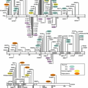
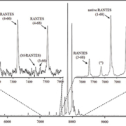
In the mass spectra, several RANTES proteoforms can be detected. As shown in Figure 1, most abundant are signals representing full-length, native RANTES (1-68) and met-RANTES, along with the N-terminally cleaved RANTES proteoforms (3-68) [MW=7,663.7; missing the ‘SP’ N-terminal dipeptide, product of dipeptidyl peptidase IV (DPP IV) enzyme cleavage] and (4-68) (MW=7,500.6; missing ‘SPY’ N-terminal tripeptide). RANTES proteoforms missing N-terminal tripeptide and C-terminal dipeptide, (4-66) (MW=7,282.3) completed the dominant signals (Figure 1, top right inlet). Additional RANTES proteoforms were identified, in lower abundance and frequency: (7-66) (MW=6993.1; missing six N-terminal and two C-terminal amino acids), (4-64) (MW=7040.1; missing three N-terminal and four C-terminal amino acids), (4-65) (MW=7153.2; missing three N- and three C-terminal amino acids) and (3-66) (MW=7445.5; missing two N- and two C-terminal amino acids). The signal labelled M-RANTES with MW=7413.5 has multiple N- and C-terminal truncation possibilities, and has not been specifically assigned. The assignation of these signals was done using the observed m/z values and the program Paws, and was in accordance with previously published qualitative results [16].
All identified RANTES proteoforms were quantified using an eight-point standard curve, in the range from 1.56 to 200 ng/mL. The standard curve was constructed from the ratio of the peak intensities of the RANTES standard and the met-RANTES IRS (y-axis) versus the RANTES standard concentration (x-axis). For the analytical samples, first, the RANTES/met-RANTES peak intensity ratios for each proteoform were determined and summed up. Using the generated standard curve equation, these ratios were used to determine the total RANTES concentration in the analysed plasma sample. Then, the concentration of the individual RANTES proteoforms was calculated based on their percentage of the total RANTES. The assay was validated through several standard procedures. The intra- and inter-assay precision experiments yielded coefficients of variation of <10%. Linearity and spiking-recovery experiments produced results between 92 and 112% (observed vs expected concentration). In a final test, the results of the RANTES MSIA were compared with those obtained with commercially available ELISA using Altman–Bland plot. A good correlation, with slight positive bias (11.3%) was obtained with the native RANTES [17].
The developed MSIA for RANTES proteoforms was applied to a cohort of 297 human plasma samples. The analyses were performed on an automated platform, which enabled for a high-throughput analysis of 96 samples in a single run. Among the samples, we were able to determine the concentration and frequency of 11 RANTES proteoforms (Figure 2). The total average concentration of RANTES was found to be 44.9 ng/ml (2.15–163 ng/mL). In majority of samples, the main proteoform was the full-length, native RANTES [c(RANTES(1-68))avg=37.4 ng/mL; 1.92–132 ng/mL], followed by RANTES (3-68), [c(RANTES(3-68))avg =6.64 ng/mL; 0.138–34.4 ng/mL]. The other truncated RANTES proteoforms were present in variable frequencies in the samples, albeit at much lower concentrations (<10% of the total RANTES). Figure 2 summarizes the distribution and frequency of all 11 RANTES proteoforms. Even though majority of RANTES proteoforms were detected in only a handful of samples and in low quantities, they should be given full attention. Cleaved proteoforms have the potential to be used as indicators of an enzymatic activity, and, in turn, of changes in the metabolic homeostasis [18]. The information that this MSIA provides puts a new perspective of RANTES quantitative analysis and can be a good starting point for looking at RANTES heterogeneity in clinical context.
Concluding remarks
The assay described above uses MALDI-TOF-MS to fully quantify RANTES proteoforms, and it is one of just a handful of such MALDI-based assays in existence today. The assay’s two-step approach is similar to that of well-established immunoassays, with the added benefit of MS detection as an enabling factor in differentiating the multiple proteoforms. The MALDI target is designed to accept the eluates from 96 tips at the same time, therefore making it high-throughput and time efficient (total time for RANTES assay is ~1 hour). The assay is performed on an automated platform, which limits the errors that can occur during assay execution. In review of previous and ongoing work, MSIA for RANTES performs well and introduces a new prospect and capacity for potential clinical applications in the field of biomarker discovery/rediscovery and diagnostics.
References
1. Strathmann FG, Hoofnagle AN. Am J Clin Pathol. 2011; 136: 609–616.
2. Agger SA, Marney LC, Hoofnagle AN. Clin Chem. 2010; 56: 1804–1813.
3. Kiernan UA, Phillips DA, Trenchevska et al. PLoS One 2011; 6: e17282.
4. Carr SA, Anderson L. Clin Chem. 2008; 54: 1749–1752.
5. Anderson NL, Anderson NG, Haines LR, et al. J Proteome Res. 2004; 3: 235–244.
6. Yocum AK, Chinnaiyan AM. Brief Funct Genomic Proteomic. 2009; 8: 145–157.
7. Nelson RW, Krone JR, Bieber AL, et al. Anal Chem. 1995; 67: 1153–1158.
8. Trenchevska O, Kamcheva E, Nedelkov D. Proteomics 2011; 11: 3633–3641.
9. Jin H, Zangar RC. Biomark Insights 2009; 4: 191–200.
10. Youn BS, Mantel C, Broxmeyer HE. Immunol Rev. 2000; 177: 150–174.
11. Lit LC, Wong CK, Tam LS, et al. Ann Rheum Dis. 2006; 65: 209–215.
12. Matter CM, Handschin C. Circulation 2007; 115: 946–948.
13. Azenshtein E, Luboshits G, Shina S, et al. Cancer Res. 2002; 62: 1093–1102.
14. Winnik S, Klingenberg R, Matter CM. Eur Heart J. 2011; 32: 393–395.
15. Kaburagi Y, Shimada Y, Nagaoka T, et al. Arch Dermatol Res. 2001; 293: 350–355.
16. Oran PE, Sherma ND, Borges CR, et al. Clin Chem. 2010; 56: 1432–1441.
17. Trenchevska O, Sherma ND, et al. J Proteomics 2014; 116C, 15–23.
18. Lim JK, Lu W, Hartley O, et al. J Leukoc Biol. 2006; 80: 1395–1404.
The authors
Olgica Trenchevska*1, Nisha D. Sherma1, Peter D. Reaven2, Randall W. Nelson1, Dobrin Nedelkov1
1Molecular Biomarkers, The Biodesign Institute at Arizona State University, Tempe, AZ, USA
2Phoenix Veterans Affairs Health Care System, Phoenix, AZ, USA
*Corresponding author
E-mail:
olgica.trenchevska@asu.edu
Point-of-care testing (POCT or POC testing) describes diagnostic tests which are performed at or physically close to a patient. This distinguishes POCT from traditional testing, which involves extracting specimens from a patient and transporting them to a laboratory for analysis. Settings for POC tests range from in-hospital bed sites and primary care offices to patient homes.
Over a half century of use
The POCT era is considered to have begun in 1962, after development of a system that measured blood glucose levels during cardiovascular surgery. The year 1977 saw the US launch of the first POC test for application wholly outside a hospital – the so-called ‘epf’ rapid pregnancy test.
POCT is increasingly used to diagnose and manage a range of diseases, from chronic conditions such as diabetes to acute coronary syndrome. One of the latest additions is a genetic test – CYP2C 19*2 allele for anti-platelet therapy.
Common POC tests includes “blood glucose testing, blood gas and electrolytes analysis, rapid coagulation testing, rapid cardiac markers diagnostics, drugs of abuse screening, urine strips testing, pregnancy testing, fecal occult blood analysis, food pathogens screening, hemoglobin diagnostics, infectious disease testing and cholesterol screening.” Nevertheless, just three tests – urinalysis by dipstick, blood glucose and urine pregnancy – are believed to account for the majority of POCT.
Turnaround time key to POCT appeal
The principal objective of POC testing is to reduce turnaround time (TAT) – a reference to the duration between a test and the obtaining of results which aid in making clinical decisions. In the past, such a process was unavoidable because of the sophistication and size of equipment required for the vast majority of medical diagnostic tests. However, technology developments have since made it possible to perform a growing number of tests outside of the laboratory.
Product miniaturization
Since the late 1980s, one of the key drivers of POCT has been product miniaturization with dedicated onboard integrated circuits. As described in a recent book on biomedical engineering, increasingly sophisticated microdevices have made it feasible to diagnose disease at point-of-care. These include “microfilters, microchannels, microarrays, micropumps, microvalves and microelectronics”, with their mechanical and electrical components “integrated onto chips to analyse and control biological objects at the microscale.” The authors list the key advantages offered by miniaturizing diagnostic tests as compared to centralized laboratory testing: portability, small size and low power consumption, simpler operation, smaller reagent volumes, faster analysis, parallel analysis, and functional integration of multiple devices.
Healthcare reforms drive POCT
Healthcare reforms have also driven POCT demand.
Spending controls and hospital mergers have led to shorter stays and faster patient turnaround. There have been growing demand for tests in outpatient clinics and patient homes. Test results have been needed quickly, not only for reasons of clinical urgency but also to ease patient waiting lists and reduce backlogs in emergency departments. Accompanying this has been the closure of several large central laboratories, which have further enhanced demand for POCT.
Making a case
The case for POCT has grown with time. In 2004, it was associated with a significant reduction in the time to treatment initiation and a shorter length of stay. More recently, a POCT cardiac marker screening stage at six UK hospitals led to a marked increase in the percentage of successful home discharges.
Such breakthroughs will increase as POCT use grows further, and as the tests become more sophisticated.
Early POC tests were based on the simple transfer of traditional methods from a central laboratory, accompanied by their downscaling to smaller platforms.
Subsequently, unique and innovative assays were designed specifically for POCT (such as the rapid streptococcal antigen test). Wide arrays of POCT-specific analytic methods have also been developed, ranging from simple (such as pH paper for assessing amniotic fluid) to the ultra-sophisticated (for example, thromboelastogram for intraoperative coagulation assessment).
Contemporary POCT systems are usually based on test kits and portable, often handheld, instruments. Many tests are realized as easy-to-use membrane-based trips, often enclosed by a plastic cassette. This requires only a single drop of whole blood, urine or saliva, and they can be performed and interpreted by any general physician within minutes.
Hospital emergency departments
Given its time-sensitive relevance, one of the fastest growing users of POCT have been hospital emergency departments (EDs).
In 2008, a study in ‘Academic Emergency Medicine’ simulated the impact of reduced turnaround times and established grounds for a “compelling improvement in ED efficiency.” Though its authors concluded that specific outcomes such as the length of stay and throughput in the emergency department warranted further investigation, they categorically recommended POCTs as a means to improve turnaround time.
Over recent years, favourable perspectives on POC tests in the ED have strengthened. At the end of last year, a study in ‘Critical Care’ found POCT increased the number of patients discharged in a timely manner, expedited triage of urgent but non-emergency patients, and decrease delays to treatment initiation. The study quantitatively assessed several conditions such as acute coronary syndrome, venous thromboembolic disease, severe sepsis and stroke, and concluded that POCT, when used effectively, “may alleviate the negative impacts of overcrowding on the safety, effectiveness, and person-centeredness of care in the ED.”
Other POCT users include ICUs as well as endocrinology, cardiology, gastroenterology and hematology.
Primary care remains principal user
The bulk of POC tests are however conducted by primary care physicians.
In 2014, the ‘British Medical Journal’ published the findings of the first-ever survey of POCT use by primary care physicians in five countries (Australia, Belgium, the Netherlands, the UK and the USA). The study found that blood glucose, urine pregnancy and urine leukocytes or nitrite were the most frequently used POC tests. Overall, more respondents in the UK and the USA reported using POC tests than respondents in the other countries. The widest gap in use of POC test was for fecal occult blood, used by 83% of US doctors against only 2–18% of primary care clinicians in the other countries.
One of the key findings of the ‘British Medical Journal’ study, however, was that there was an unmet need for new POC tests. Included here were tests for D-dimer, troponin, chlamydia, gonorrhea, B-type natriuretic peptide, CRP, glycated hemoglobin, white cell count and hemoglobin, which were desired by more than half of respondents across all the five countries.
Fast growing market
Over the past two-and-a-half decades, the availability and use of POCT has steadily increased. By 2012, nearly 100 companies worldwide were developing, manufacturing or marketing POC tests. One study, cited by the National Institutes of Health in the US, places POCT sales in 2011 at about $15 billion (€13.5 billiion). Of this figure, the US accounted for a share of 55%, Europe for 30% and Asia for 12%. The market is projected to show compound annual growth of 4% to reach $18 billion (€16.2 billion) by 2016.
Further growth in the use of POCT is expected to be driven by increases in accuracy, reliability and convenience. Alongside, one of the biggest catalysts for increased POCT use may consist of quality standards.
The quality challenge
Issues about POCT quality continue to vex experts. Variability in the interpretation of POC test results is a widespread concern, given differences in the education and experience of staff who conduct the tests. In addition, POCT results may also not be comparable across sites (e.g. when patients travel) and differences in specimen types (serum, plasma or whole blood) can impact on results – as compared to those from a traditional central laboratory.
In a laboratory setting, analytical quality is usually assessed by QC (quality control) and QA (quality assurance) procedures. Their aim is “to monitor the stability of the analytical measurement system and to alert the operator to a change in stability”… “that may lead to a medically important error.” While these processes serve a laboratory well, it is unclear whether these processes are relevant, transferable and practical for monitoring quality on POCT devices.
Regulators and POCT in the US and the EU
Future developments are expected to be driven by regulatory bodies.
In the US, CLIA88 (Clinical Laboratory Improvement Amendments of 1988) provided a major impetus for growth in POCT. The rules, published in 1992, expanded the definition of ‘laboratory’ to include any site where a clinical laboratory test occurred (including a patient’s bedside or clinic) and specified quality standards for personnel, patient test management and quality.
One of CLIA88’s biggest contributions to POCT growth was to define tests by complexity (waived, moderate complexity and high complexity control), with minimal quality assurance for the waived category.
CLIA88 has been followed by US federal and state regulations, along with accreditation standards developed by the College of American Pathologists and The Joint Commission. These have established POCT performance guidelines and provided strong incentives to ensure the quality of testing.
In Europe, POCT devices are regulated under the 1998 European Directive 98/79/EC on in vitro diagnostic medical devices, although the term itself is not specifically mentioned. There have since been several amendments, most recently in 2011 (2011/100/EU), as well as standards based on the Directive’s framework.
However, at the European level, specific coverage of POCT is referred by international standard ISO 22870:2006, used in conjunction with ISO 15189 which covers competence and quality in medical laboratories. It is important to note that patient self-testing in a home or community setting is not covered by ISO standards.
The role of ICT
The role of ICT in driving the growth of POCT is also likely to become crucial. In the late 1990s, there were concerns that POCT implementation, especially in the real-time critical care context, was accompanied by little understanding of its information technology requirements.
However, the situation has since changed dramatically, especially as ICT is seen as the only appropriate interface between POC test results and computerized patient records – seen as the means to restructure clinical care pathways.
ICT is also accepted as the best means to standardize care protocols. In 2012, a study found that the impact of point-of-care panel assessment on successful discharge and costs varied markedly from one hospital to another and that outcomes depended on local protocols, staff practices and available facilities. In effect, the study highlighted the importance of optimizing clinical pathways to derive maximum benefit from the reduced turnaround times provided by POCT.
When founding the company GONOTEC GmbH in 1979, electronics engineer Harald Göritz and chemist Klaus Noack could not possibly imagine that their target to develop, produce and market analytical measuring instruments for medical and chemical application would be as successful as it turned out to be. Both founders of the company could already look back to decades of experience in this field.
It all started with a cryoscopic osmometer for clinical application: the OSMOMAT 030. At the first exhibitions he visited, Klaus Noack was surprised by the high level of interest generated by the instrument. There was no other way than to expand production to cope with the increasing demand for this osmometer, which offered a complete ease of handling, previously unattained, resulting in a growing number of sales.
Inspired by this success, a new osmometer was developed by Klaus Noack to complete the osmometer line for medical application: the colloid osmometer OSMOMAT 050. Even though the market for this instrument, basically used in intensive care units, was not as big as for the OSMOMAT 030, the product also contributed to the further success of GONOTEC GmbH.
In the middle of the 80’s, development started on a new range of instruments, namely the chemical osmometers. The aim was the development of instruments for the determination of molar masses based on osmotic parameter for chemical application that also offer easy handling for the user.
The three osmometers, vapor pressure osmometer OSMOMAT 070, membrane osmometer OSMOMAT 090 and cryoscopic osmometer OSMOMAT 010 complement one another due to their different measuring methods in the determination of the range of molar masses up to 2,000,000 Dalton.
In 2001 the general management of GONOTEC was taken over by Jan Celinsek, who worked already with GONOTEC since 1991.
In 2003 a new model in the osmometer family was launched into the market: the OSMOMAT auto, which is also characterized by extreme reliability and easy handling, thus fitting perfectly into the already well known GONOTEC osmometer line.
In 2009 GONOTEC moved to new premises with lots of space for new ideas! In the same year, the chloridmeter CM20 was launched, followed in 2013 by the next generation of osmometers: the OSMOMAT 3000 and the OSMOMAT 3000 basic, both replacing the well known OSMOMAT 030.
To this day, GONOTEC is no large, anonymous concern but still a medium-sized, private company, owned by Klaus Noack, the founder. However, we became a global player with customers in more than 60 countries.
One of the most valuable resources GONOTEC always had was its permanent staff. Once people start working for GONOTEC they stay with the company as they are proud of their work. The same applies to the numerous number of dealers all over the world. The cooperation between the agents and GONOTEC is like in a family; constant trainings at the company headquarters improve this special relationship between agent and manufacturer. GONOTEC products do deserve the description “Made in Germany”, as the whole production is in one location. It is easy for external persons visiting the company to see an osmometer being manufactured from the very beginning to its finishing and perfect functioning.
Since GONOTEC was able to export the company’s philosophy by means of the highest quality standards and competence as well as constant assistance to customers and agents, it is looking optimistically into the future. Our company’s philosophy is a promise to all our customers and potential customers.
The anti-TNF therapies infliximab and adalimumab have revolutionized the treatment of inflammatory bowel disease, being very effective in many patients. Some patients experience problems such as loss of response, which is associated with production of antibodies to the therapy. Measuring trough drug and antibody concentrations may direct patient management in future.
by Dr Mandy Perry, Dr Tim McDonald, Adrian Cudmore, Dr Tariq Ahmad
Ulcerative colitis (UC) and Crohn’s disease (CD) are relapsing and remitting inflammatory disorders of the gastrointestinal (GI) tract. Recently published data suggests that as many as 620 000 people in the UK could have these inflammatory bowel diseases (IBDs). Both conditions can produce symptoms of urgent and frequent diarrhea, rectal bleeding, pain, profound fatigue and malaise. In some patients, there is an associated inflammation of the joints, skin, liver or eyes. Malnutrition and weight loss are common, particularly in CD. These conditions can cause considerable disruption to education, working, social and family life. There is currently no cure. Drugs to suppress the immune system are the mainstay of medical management, and first line treatment typically includes corticosteroids, with immunmodulators such as azathioprine, mercaptopurine or methotrexate used for patients with steroid-dependent disease. However, 30% of patients either fail to respond, or are intolerant, to these drugs and will then be considered for biological therapies or surgery. More than half of patients with CD and about 20–30% of patients with UC will require surgery at some point. The anti-TNF agents infliximab and adalimumab have revolutionized treatment of IBD, and are an effective alternative to surgery, leading to complete remission in many patients [1].
NICE has published guidelines for the use of anti-TNF agents for CD [2] and UC [3]. These drugs include the monoclonal anti-TNF drugs infliximab (includes the original product – Remicade, and biosimilar infliximab Remsima and Inflectra) and adalimumab (Humira). TNF is a cytokine involved in systemic inflammation and the anti-TNF drugs bind to and inactivate TNF, thereby halting the immune cascade and reducing inflammation. Infliximab is a mouse–human chimeric anti-human TNF antibody which is administered by intravenous infusion with a typical induction course of therapy at weeks 0, 2, 6 and then 8-weekly maintenance dose. Adalimumab is a fully human anti-human TNF antibody, and is administered by subcutaneous injection every 2 weeks. For some patients this is a more convenient option, as the subcutaneous rather than intravenous administration means that frequent hospital appointments are not required. Both infliximab and adalimumab are expensive treatments, typically costing in excess of £10,000 per annum. The 2015 introduction of biosimilar infliximab preparations has significantly reduced the price of therapy.
Some patients have an excellent response to anti-TNF treatment, managing to obtain complete remission of CD and mucosal healing. However, a proportion of patients do not respond well to anti-TNF therapy [4], and there are three principal problems:
The etiology of these problems is unknown, although the following causes have been indicated [1]:
Approximately 25% of patients will develop antibodies to infliximab and adalimumab drugs within 12 months of treatment initiation. The clinical importance of such antibodies is not completely understood. It is hypothesized that anti-drug antibodies may alter the action of the drug (i.e. neutralizing) and/or increase the drug clearance (i.e. non-neutralizing). Antibodies may be transient (and may be ‘overcome’ by increasing the concentration of drug), or persistent (and intolerant of drug escalation) [6, 7].
Measuring drug and anti-drug antibodies may enable problems such as ADR, PNR and LOR to be further understood, and may assist clinicians in the management of these problems. Possible interventions include escalating the dose of anti-TNF therapy, adding in an additional drug (e.g. immunomodulator or steroid), switching to an alternative anti-TNF therapy or switching to a non-TNF biologic.
For infliximab, several algorithms for patient management have been developed using drug and antibody levels [8, 9]. Several different assays, using different therapeutic ranges have been employed as part of these algorithms, making comparison difficult. The widely quoted TAXIT (Trough level Adapted infliXImab Treatment) trial uses a therapeutic range for infliximab of 3–7 mg/L [8], whereas work by Steenholdt uses 5–10 mg/L [9]. The different technologies used to measure drug and anti-drug antibodies, include ELISA (enzyme-linked immunosorbent assay), HMSA (homogeneous mobility shift assay) [10], radioimmunoassay, and a functional cell-based reporter gene assay [11]. There is poor agreement between the drug assays, as there is neither gold standard material, nor a reference method available.
Clinicians and laboratories should also be aware that there is considerable variation in what is being measured for the antibody assays. For example, the ELISA antibody assays either measure free (i.e. only those antibodies which are not bound to drug in the patient serum) or total antibodies (i.e. bound and unbound to drug). The functional cell-based assay is different again, as it is designed to detect only those antibodies which prevent infliximab from binding to TNF and therefore may not detect antibodies that are postulated to alter the drug clearance only. Although anti-TNF drug and antibody testing shows promise, there is not yet sufficient cost-effective data, nor diagnostic algorithms, for widespread adoption across the NHS. It seems likely that use in the setting of loss of response will enter clinical practice first and may allow cost savings by avoiding dose escalation in patients with high levels of antibodies.
The Personalized Anti-TNF Therapy in Crohn’s disease (PANTS) study is a prospective, observational study for which anti-TNF naïve patients aged 6 and over are eligible. The study aims to investigation the clinical, serological and genetic factors that determine PNR, LOR and ADR to anti-TNF drugs in patients with active luminal Crohn’s disease. The study is recruiting from over 110 UK hospitals currently participating in the UK Inflammatory Bowel Disease Genetics Consortium pharmacogenetic programme. While attending routine clinical appointments, additional information and samples are collected for the PANTS project. This includes the Harvey Bradshaw index (HBI, a scoring system which classifies recent disease in terms of symptoms), blood for DNA, RNA, CRP (C-reactive protein), anti-TNF drug and antibody levels and stool samples for calprotectin. Analysis of CRP, calprotectin and anti-TNF alpha drug and antibody levels is undertaken at the Central Laboratory at Exeter Blood Sciences Laboratory, where a biobank of additional serum aliquots is being constructed. Infliximab and adalimumab drug levels, total anti-infliximab antibody and total anti-adalimumab antibody are measured by ELISA technology (Immundiagnostik), using a liquid handling robot (DS2, DYNEX Technologies) [12]. Biochemical data is uploaded onto a bespoke web-based database that is also used to store the clinical information.
Examples of data for two patients from the PANTS study are shown in Table 1. Table 1A is data from a patient who is prescribed infliximab. Week 0 shows baseline data before treatment with infliximab. The calprotectin is raised, indicating active inflammation, and this is mirrored with the CRP and Harvey Bradshaw Index (HBI score of <5 indicates remission; 5–7 mild disease, 8–16 moderate disease and >16 severe disease). By week 14, the calprotectin has decreased substantially, and the CRP and HBI have decreased to normal values at week 2. Until the end of the timeframe (week 126), the patient continues to have a normal calprotectin, CRP and HBI. The drug level concentration in the maintenance phase is between 3–14 mg/L and the patient remains negative for anti-drug antibodies (i.e. <10 AU/mL). Table 1B shows data from a pediatric patient who is prescribed infliximab. The PCDAI (Pediatric Crohn’s Disease Activity Index) is used in place of the HBI. When infliximab naïve (week 0), the patient had a raised CRP, calprotectin and PCDAI (<10 remission; 10–29 mild disease; 30–39 moderate disease; >40 severe disease). Upon treatment with infliximab there was initially a good response, shown by the decrease in CRP and PCDAI. At week 22, the patient became positive for anti-drug antibodies and the trough drug concentration became undetectable. At week 26, the patient had clinical loss of response and underwent surgery. Knowledge of the patient’s drug and antibody levels helps with clinical management in the setting of loss of response, such as in this case. Dose escalation is likely to be futile and costly in patients with high antibody titres. Switching to an alternative anti-TNF might provide transient benefit, although patients who form antibodies to one anti-TNF are likely to form antibodies to the second and subsequent agents in this class.
The anti-TNF drugs infliximab and adalimumab are effective treatment for CD in many patients. However, LOR, PNR and ADR are significant problems, and it is so far unclear as to how these patients should be best managed. Measuring drug and antibody concentrations may allow for diagnostic algorithms to be produced. The clinical and cost effectiveness of therapeutic monitoring of TNF inhibitors using ELISA technology is currently being evaluated by NICE [13]. It is anticipated that data from the PANTS study will directly inform such algorithms and guidelines, and contribute to an evidence based medicine approach for management of CD patients who are prescribed anti-TNF therapy.
References
1. Vande Casteele N, Feagan BG, Gils A, et al. Therapeutic drug monitoring in inflammatory bowel disease: current state and future perspectives. Curr Gastroenterol Rep. 2014; 16: 378.
2. NICE technology appraisal guidance [TA187]. Infliximab (review) and adalimumab for the treatment of Crohn’s disease. NICE 2010. (https://www.nice.org.uk/guidance/ta187/chapter/1-guidance).
3. NICE technology appraisal guidance [TA329]. Infliximab, adalimumab and golimumab for treating moderately to severely active ulcerative colitis after the failure of conventional therapy (including a review of TA140 and TA262). NICE 2015. (https://www.nice.org.uk/guidance/ta329)
4. Nielsen OH, Seidelin JB, Munck LK, Rogler G. Use of biological molecules in the treatment of inflammatory bowel disease. J Int Med. 2011; 270: 15–28.
5. Vande Casteele N, Ballet V, Van Assche G, et al. Early serial trough and antidrug antibody level measurements predict clinical outcome of infliximab and adalimumab treatment. Gut 2012; 61: 321.
6. Hanauer S, Feagan B, Lichtenstein G, et al. Maintenance infliximab for Crohn’s disease: the ACCENT I randomised trial. Lancet 2002; 359: 1541–1549.
7. Cornillie F, Hanauer B, Diamond R, et al. Postinduction serum infliximab trough level and decrease of C-reactive protein level are associated with durable sustained response to infliximab: a retrospective analysis of the ACCENT I trial. Gut 2014; 63; 1721–1727.
8. Vande Casteele N, Ferrante M, Van Assche G, et al. Trough concentrations of infliximab guide dosing for patients with inflammatory bowel disease. Gastroenterology 2015; 148: 1320-1329.
9. Steenholdt C, Brynskov J, Thomsen OØ, et al. Individualised therapy is more cost-effective than dose intensification in patients with Crohn’s disease who lose response to anti-TNF treatment: a randomised, controlled trial. Gut 2014; 63: 919–927.
10. Wang SL, Ohrmund L, Hauenstein S, et al. Development and validation of a homogeneous mobility shift assay for the measurement of infliximab and antibodies-to-infliximab levels in patient serum. J Immunol Methods 2012; 382: 177–188.
11. Lallemand C, Kavrochorianou N, Steenholdt C, et al. Reporter gene assay for the quantification of the activity and neutralizing antibody response to TNFα antagonists. J Immunol Methods 2011; 373: 229–239.
12. Perry M, Bewshea C, Brown R, et al. Infliximab and adalimumab are stable in whole blood clotted samples for seven days at room temperature. Ann Clin Biochem. 2015; doi: 10.1177/0004563215580001.
13. NICE. Crohn’s disease – Tests for therapeutic monitoring of TNF inhibitors (LISA-TRACKER ELISA kits, TNFa-Blocker ELISA kits, and Promonitor ELISA kits). Anticipated publication date: December 2015. (https://www.nice.org.uk/guidance/indevelopment/gid-dt24/consultation/crohns-disease-tests-for-therapeutic-monitoring-of-tnf-inhibitors-lisatracker-elisa-kits-tnfablocker-elisa-kits-and-promonitor-elisa-kits-consultation)
The authors
Mandy Perry*1 PhD, Tim McDonald1 FRCPath. PhD, Adrian Cudmore1, Tariq Ahmad2 MB ChB, DPhil, MRCP(UK)
1Department of Blood Sciences, Royal Devon and Exeter NHS Foundation Trust, Exeter, UK
2IBD Pharmacogenetics Research, University of Exeter, Exeter, UK
*Corresponding author
E-mail: mandy.perry@nhs.net
November 2024
The leading international magazine for Clinical laboratory Equipment for everyone in the Vitro diagnostics
Beukenlaan 137
5616 VD Eindhoven
The Netherlands
+31 85064 55 82
info@clinlabint.com
PanGlobal Media is not responsible for any error or omission that might occur in the electronic display of product or company data.
This site uses cookies. By continuing to browse the site, you are agreeing to our use of cookies.
Accept settingsHide notification onlyCookie settingsWe may ask you to place cookies on your device. We use cookies to let us know when you visit our websites, how you interact with us, to enrich your user experience and to customise your relationship with our website.
Click on the different sections for more information. You can also change some of your preferences. Please note that blocking some types of cookies may affect your experience on our websites and the services we can provide.
These cookies are strictly necessary to provide you with services available through our website and to use some of its features.
Because these cookies are strictly necessary to provide the website, refusing them will affect the functioning of our site. You can always block or delete cookies by changing your browser settings and block all cookies on this website forcibly. But this will always ask you to accept/refuse cookies when you visit our site again.
We fully respect if you want to refuse cookies, but to avoid asking you each time again to kindly allow us to store a cookie for that purpose. You are always free to unsubscribe or other cookies to get a better experience. If you refuse cookies, we will delete all cookies set in our domain.
We provide you with a list of cookies stored on your computer in our domain, so that you can check what we have stored. For security reasons, we cannot display or modify cookies from other domains. You can check these in your browser's security settings.
.These cookies collect information that is used in aggregate form to help us understand how our website is used or how effective our marketing campaigns are, or to help us customise our website and application for you to improve your experience.
If you do not want us to track your visit to our site, you can disable this in your browser here:
.
We also use various external services such as Google Webfonts, Google Maps and external video providers. Since these providers may collect personal data such as your IP address, you can block them here. Please note that this may significantly reduce the functionality and appearance of our site. Changes will only be effective once you reload the page
Google Webfont Settings:
Google Maps Settings:
Google reCaptcha settings:
Vimeo and Youtube videos embedding:
.U kunt meer lezen over onze cookies en privacy-instellingen op onze Privacybeleid-pagina.
Privacy policy